|
Case Report
Perioperative hypersensitivity to a vaginal cuff hemostatic agent in the setting of alpha-gal syndrome
1 Resident, Department of Obstetrics and Gynecology, Oklahoma State University Medical Center, Tulsa, OK, USA
2 Gynecologist, Department of Obstetrics and Gynecology, Cherokee Nation W. W. Hastings Hospital, Tahlequah, OK, USA
3 Dean, Oklahoma State University College of Osteopathic Medicine at the Cherokee Nation, Tahlequah, OK, USA
4 Medical student, Oklahoma State University College of Osteopathic Medicine at the Cherokee Nation, Tahlequah, OK, USA
5 Clinical associate professor, Oklahoma State University College of Osteopathic Medicine at the Cherokee Nation, Tahlequah, OK, USA
Address correspondence to:
Bridget Botsford
717 S Houston Avenue #200, Tulsa, OK 74127,
USA
Message to Corresponding Author
Article ID: 100047G06BB2026
Access full text article on other devices

Access PDF of article on other devices


How to cite this article
Botsford B, Dorsett T, Bray N, Shed A, Jones A. Perioperative hypersensitivity to a vaginal cuff hemostatic agent in the setting of alpha-gal syndrome. Edorium J Gynecol Obstet 2026;10(2):15–23.ABSTRACT
Introduction: Alpha-gal syndrome (AGS) is an increasingly recognized immunoglobulin E (IgE)-mediated hypersensitivity disorder to mammalian-derived products that carries serious surgical implications if not considered perioperatively. As hemostatic agents are frequently utilized during gynecologic surgery, surgeons must be familiar with their components and potential adverse effects. Delayed hypersensitivity reactions (DHRs) to animal-derived surgical materials remain an underexplored area. Heightened surgeon awareness of appropriate hemostatic agent selection is critical, particularly in patients with AGS.
Case Report: We present the perioperative course of a 42-year-old woman with known AGS who underwent an elective hysterectomy for abnormal uterine bleeding and dysmenorrhea. Intraoperatively, a bovine-derived hemostatic agent was applied to the vaginal cuff. Her postoperative course was complicated by a delayed presentation of pelvic hematoma, abscess formation, vaginal cuff dehiscence, and necrosis, requiring three additional surgeries. Notably, this patient demonstrated persistent eosinophilia without leukocytosis despite pathologically confirmed pelvic abscesses—an atypical finding suggestive of an immune-mediated process rather than isolated infection.
Conclusion: The clinical course is consistent with a DHR superimposed on secondary infection following intraoperative exposure to a bovine-derived hemostatic agent in a patient with known AGS. This case highlights the potential severity, adverse outcomes, protracted hospital course, and resource utilization associated with inadvertent exposure to animal-derived surgical materials in patients with AGS. Delayed hypersensitivity reactions may not be immediately recognized, so surgeons should maintain heightened vigilance when operating on patients with known AGS and explore alternative hemostatic agents to prevent avoidable adverse outcomes.
Keywords: Alpha-gal syndrome, Delayed hypersensitivity, Gynecologic surgery, Hemostatic agents
INTRODUCTION
Alpha-gal syndrome (AGS) is an immunoglobulin E (IgE)-mediated allergy to mammalian meat and mammalian-derived products as a result of Lone Star tick (Amblyomma americanum) bites [1]. During feeding, the tick transfers an oligosaccharide, galactose-α-1,3-galactose (alpha-gal), into the dermis, initiating the development of IgE antibodies [2]. Alpha-gal is present in mammalian tissues. For individuals who have not been sensitized, dietary exposure to the alpha-gal in red meat is not harmful. When an individual has been sensitized and they are subsequently exposed to mammalian-derived antigens, this leads to the activation of basophils and mast cells, ultimately inciting the release of histamine [3]. Depending on the affected individual’s gastrointestinal transport time, the presentation of symptoms varies though is typically delayed, occurring approximately 2–6 hours after the time of exposure. Symptoms can range from mild (e.g., cough, indigestion, abdominal pain, and urticaria) to severe (e.g., angioedema, difficulty breathing, and anaphylaxis) [4].
This delayed presentation distinguishes AGS from most immediate food allergies and can complicate the diagnosis of AGS in clinical settings. The diagnosis of AGS is made based on evidence of AGS sensitization and having a compatible clinical history [5]. While there is not a well-established alpha-gal IgE level to diagnose, most reports refer to a value of >0.1 IU/mL as a positive result [5]. In addition to red meat, alpha-gal is also found in mammalian-derived products, including gelatin- and glycerin-containing products. While the association between AGS and reactions to mammalian meat has been well described, comparatively little attention has been given to hypersensitivity reactions involving mammalian-derived intraoperative materials, particularly delayed reactions.
The prevalence of AGS in the United States is unknown due to the lack of a national surveillance system. The Centers for Disease Control and Prevention (CDC) evaluated the geographic distribution of the 110,000 suspected cases between 2010 and 2022 and identified the highest prevalence in the mid-Atlantic, southern and mid-western United States [6]. Binder et al. estimated approximately 2–5 cases per 100,000 persons in these areas, acknowledging that methodologic limitations may have led to underestimation of true prevalence [7]. The prevalence of AGS has increased substantially over all demographics and continues to rise [8]. A study by Ross et al. used predictive modeling to evaluate expected growth of cases by the year 2030. Black and Hispanic populations were projected to be disproportionately affected with expected incidences of 57% and 72%, respectively [8]. The suspected current prevalence of AGS and projected rise highlights the necessity for awareness of this syndrome.
When patients undergo surgical intervention, it is important to consider perioperative planning. A thorough review of the patient’s complete medical history and allergies are accounted for when deciding on preoperative antibiotics, anesthetic agents, operative materials, and postoperative medications. The patient’s unique history should tailor their perioperative planning as medicine is extraordinarily personalized. Alpha-gal syndrome is underrecognized and poorly understood by providers. In one report, a survey was administered to providers and cited that 42% were unaware of the syndrome and 35% endorsed a lack of confidence in diagnosing or managing these patients [9]. Alpha-gal syndrome should be treated with similar caution to other allergies, and surgeon education is of utmost importance to avoid potential life-threatening complications.
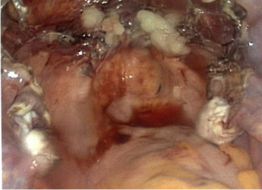
We present a 42-year-old woman with known AGS whose elective hysterectomy was complicated by a protracted course requiring three additional surgeries, suspected to be attributable to exposure to a bovine-derived hemostatic agent (Figure 1). Current literature describes immediate hypersensitivity reactions to mammalian-derived surgical materials and delayed reactions following ingestion of mammalian meat but lacks documentation of delayed reactions to mammalian-derived surgical materials. The objective of this case report is to describe the complicated perioperative course of a patient with known AGS, explain the suspected delayed reaction that occurred, raise awareness of this syndrome, and review perioperative considerations.
CASE REPORT
We present a 42-year-old Native American woman with known AGS who presented for elective total vaginal hysterectomy with bilateral salpingectomy (TVH-BS) for abnormal uterine bleeding and dysmenorrhea. Her surgical history included two cesarean sections and two prior supraumbilical hernia repairs.
She had been diagnosed with AGS three years prior, following detection of elevated alpha-gal IgE levels during evaluation for new-onset fatigue, bloating, and night sweats. This patient was diagnosed by an IgE level of 0.22 IU/mL and improvement of symptoms with elimination of triggering foods. Her AGS diagnosis was managed by her primary care physician with prescriptions for cetirizine and hydroxyzine alongside advice to avoid potential triggers. The patient was not evaluated by an allergist or immunologist.
Initial Surgery
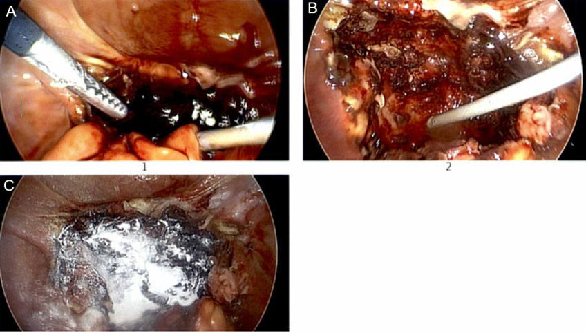
The TVH-BS proceeded without complication and was followed by diagnostic laparoscopy to further evaluate the ovaries in the setting of dysmenorrhea. A unilateral ovarian cystectomy was performed. The vaginal cuff was closed using a synthetic suture. During the surgery, a hemostatic agent composed of a bovine-derived gelatin matrix and a thrombin component was applied to the vaginal cuff for hemostasis (Figure 2). The patient’s initial hospital course was uncomplicated, and she was discharged once she achieved all postoperative milestones. She was sent home with prescriptions for oral tablets of Percocet, gabapentin, ibuprofen, and Zofran.
First Readmission (Postoperative Week 1)
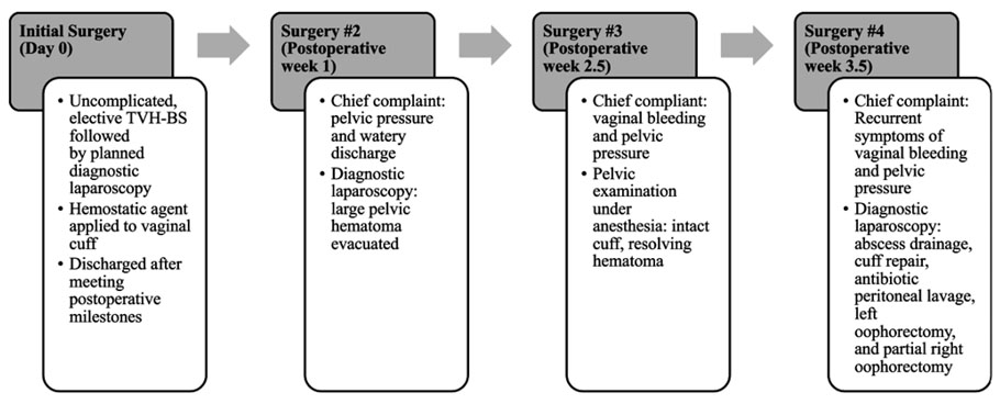
Nine days after surgery, the patient presented to the clinic for evaluation due to symptoms of pelvic pressure and clear, watery vaginal discharge. She reported that these adverse symptoms began seven days after surgery. On physical examination she had mild lower abdominal tenderness. Pelvic examination revealed an intact vaginal cuff with notable tenderness to palpation. There was suspicion for possible vaginal cuff cellulitis, but she was sent to the emergency department (ED) for further workup. In the ED, she was noted to have stable vital signs and a normal white blood cell count. A computed tomography scan of the pelvis demonstrated concern for a 7.6 cm pelvic abscess with moderate complex free fluid.
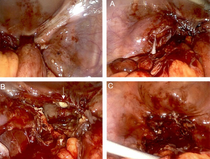
She was returned to the operating room for diagnostic laparoscopy, which revealed a large, dark-red clotted hematoma covering the vaginal cuff and the entire posterior cul-de-sac (Figure 3A). Surgical evacuation of the hematoma was performed, and there was no active bleeding appreciated underneath it. The operative report also mentioned that there were no signs of abscess or pelvic infection at that time, but raw appearing tissue was seen along the bladder and posterior peritoneum. A small area of oozing of blood was noted from the site of previous cystectomy which was cauterized. A plant-based hemostatic agent was applied to the vaginal cuff and affected ovary. The patient was discharged on postoperative day three following establishment of an AGS-compatible pain regimen (Figure 3B and Figure 3C).
Second Readmission (Postoperative Week 2.5)
The patient reported that since she was discharged home, she continued to feel a sensation of pelvic fullness. Despite this, she did not seek out evaluation until five days after being discharged home (approximately 2.5 weeks following index surgery) due to new onset vaginal bleeding. Pelvic examination under anesthesia revealed an intact vaginal cuff without drainage and a small area of supracuff fullness consistent with a resolving hematoma. She was discharged the same day.
Third Readmission (Postoperative Week 3.5)
Five days after her pelvic examination under anesthesia (approximately 3.5 weeks from index surgery), the patient again presented with vaginal bleeding and pelvic pressure. She reported that these symptoms never stopped since being discharged home. Imaging demonstrated findings consistent with multiple pelvic abscesses, largest measuring 5.4 cm × 4.7 cm in diameter. Repeat diagnostic laparoscopy was performed, with drainage of multiple pelvic abscesses, peritoneal lavage, and antibiotic irrigation. The vaginal cuff was notably friable with tissue sloughing (Figure 4A and Figure 4C). Necrotic ovarian tissue was identified, necessitating right oophorectomy and left partial oophorectomy. Vaginal cuff dehiscence was identified and repaired in the same setting with use of a different synthetic suture (Figure 4B).
The patient was initiated on Zosyn antibiotic therapy. In the following postoperative days, suspicion began to rise that the patient’s AGS may be complicating her perioperative course. Benadryl and famotidine were added to her regimen on postoperative day three with hopes of reducing vaginal cuff irritation. Her abscess culture resulted positive for Escherichia coli with sensitivity to Zosyn. Repeat imaging was performed on postoperative day five as the patient’s pelvic pain and discomfort continued. Although the abscesses decreased in size postoperatively, persistence of the collections prompted transfer to a tertiary facility for image-guided abscess drain placement by interventional radiology.
The patient underwent drain placement at the outlying facility, completed an intravenous antibiotic course and returned for a follow up clinic visit approximately two weeks later. The patient reported daily improvement in symptoms and did not return to the operating room after this intervention.
Pathologic Analysis
The right ovary and left ovarian cyst wall were submitted for pathologic evaluation and demonstrated benign ovarian tissue with acute and chronic inflammation, abscess formation, and focal granulomas with necrosis.
Laboratory Findings
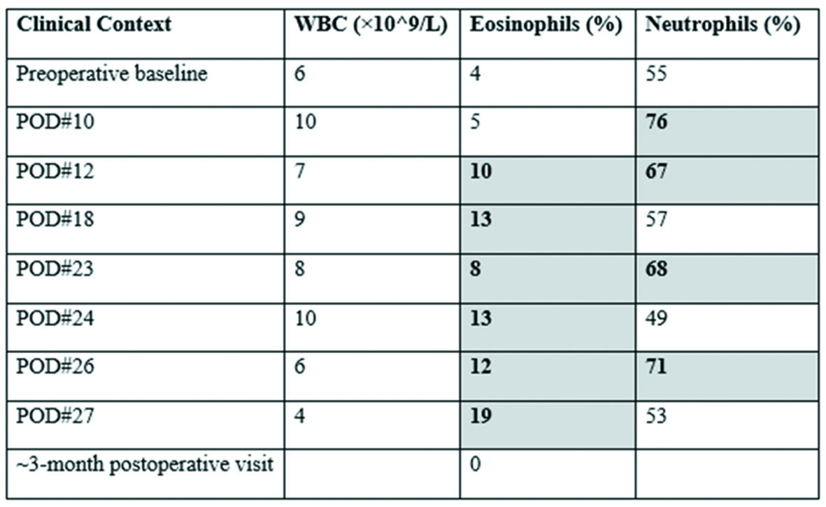
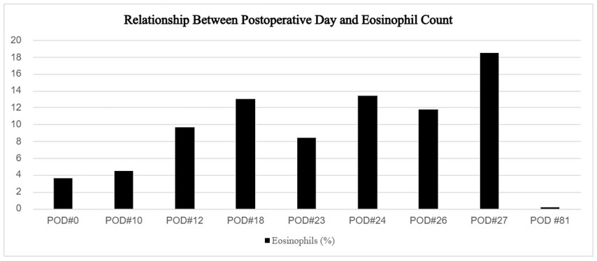
Throughout her postoperative course, the patient’s white blood cell count remained within or near normal limits despite pathologically confirmed pelvic abscesses—an atypical finding inconsistent with a purely infectious etiology (Figure 5). In contrast, persistent and progressive eosinophilia was observed across multiple time points, peaking at 19% on postoperative day 27 before resolving to 0% at an approximately three-month follow-up visit (Figure 5 and Figure 6). This pattern of recurrent eosinophilia in the absence of persistent leukocytosis raises significant concern for an underlying hypersensitivity-driven inflammatory process.
DISCUSSION
Delayed Hypersensitivity Reactions and AGS
Alpha-gal syndrome is classified as a type I hypersensitivity reaction which is IgE-mediated. First, sensitization occurs when humans develop IgE antibodies against alpha-gal which is transmitted from the initial tick bite. Upon re-exposure, the sensitized individual then absorbs alpha-gal antigen which binds to IgE on mast cells. The mast cells then degranulate, and histamine release occurs which leads to the physical manifestations of this reaction [10]. Type I hypersensitivity reactions are typically rapid immune system responses which may be seen when patients with AGS are exposed to intravenous alpha-gal [11]. It is well documented that in contrast, when patients ingest animal-derived products, this hypersensitivity reaction is instead delayed. The reasoning for this delay is not well understood and differs from the typical immediate onset with other common food allergies [11]. The leading hypothesis is that the delay relates to the time it takes the body to process glycolipids such as alpha-gal into the size of low-density lipoprotein (LDL) [12]. Interestingly, this case report demonstrated a response that was delayed past the cited 2–6 hours commonly seen after ingestion of mammalian-meat products in patients with AGS.
A DHR, or type IV hypersensitivity reaction, is a CD4+ and CD8+ T-cell-mediated immune response that typically manifests 48–72 hours following antigen exposure, though onset may extend to several weeks [13]. The type IVa subtype is classically seen in allergic contact dermatitis. There is first a sensitization phase which can take days to years. First, an antigen penetrates the epidermis of the host. Antigen presenting cells then process the antigen which migrates to regional lymph nodes [14]. The processed peptide is presented to T cells which drives clonal proliferation and differentiation [15]. Memory T cells then develop from this process. The second phase is the elicitation phase where the host is re-exposed to the same antigen and memory T cells are recruited and re-activated. Cytokine release recruits cytotoxic T cells and macrophages which release lytic enzymes and reactive oxygen species [15]. The result is inflammation and tissue damage. If the antigen cannot be cleared, granuloma formation may occur [15].
In type IV hypersensitivity reactions, the delay in the reaction is due to the time required for cell recruitment and activation. In AGS, the type I hypersensitivity reaction that is usually defined as being immediate is instead delayed, which is hypothesized to be because of delayed gastrointestinal transit time. The patient in this case report had her first readmission one week after initial exposure to a mammalian-derived hemostatic agent, and complications continued for weeks after then. This patient’s reaction is not fully explained by an expected delayed, type I hypersensitivity reaction but rather followed a much-delayed presentation, similarly to how a type IV hypersensitivity reaction may present.
Clinical Correlation
What distinguished this patient’s course was the temporal dissociation between surgical exposure and clinical deterioration, combined with a laboratory profile inconsistent with isolated bacterial infection. The absence of leukocytosis in the setting of pathologically confirmed abscesses is atypical and may indicate a hypersensitivity-driven inflammatory etiology rather than a purely infectious one. A hypothesized ongoing immune-mediated process is further supported by the persistent and recurrent eosinophilia observed throughout her admissions.
We propose that this patient’s protracted postoperative course represents an acute bacterial infection superimposed on a chronic inflammatory reaction in the context of known AGS. While hematomas and pelvic abscesses may occur in patients without AGS, the recurrent nature of her complications extending weeks beyond the index surgery, combined with persistent eosinophilia, raises concern for a chronic inflammatory substrate. The intraoperative findings of progressive tissue necrosis, vaginal cuff friability, tissue sloughing, and focal granulomas on pathologic analysis further support this hypothesis. The bovine-derived hemostatic agent applied at the time of index surgery, is known to persist in tissue for approximately 6–8 weeks—a duration sufficient to sustain prolonged mammalian antigen exposure and potentially perpetuate an ongoing inflammatory response in a sensitized host [16].
Exclusion of Other Etiologies
When reviewing this patient’s perioperative course, many additional factors were excluded prior to attributing the highest suspicion of adverse outcomes to the hemostatic agent that was utilized. When evaluating preoperative antibiotics, Ancef was administered prior to the initial surgery and is not known to contain any animal-derived molecules [17],[18]. Additionally, adverse effects would be expected to occur immediately and not delayed by days to weeks. The agents that were administered by the anesthesia team (dexmedetomidine, midazolam, fentanyl, lidocaine, propofol, rocuronium, dexamethasone, phenylephrine, fluorescein, furosemide, ketorolac, sugammadex, and meperidine) are generally considered safe in AGS [18]. Animal or plant-based glycerin may be used to hydrate propofol based on the manufacturer that is used, but even if it is animal-derived, it has not been confirmed to contain alpha-gal and is unlikely to cause an adverse reaction in patients with AGS [19].
When reviewing how the patient was prepared for each of these surgeries, the hospital utilized chlorhexidine abdominal preparation and a povidone-iodine vaginal preparation. It is documented in the patient’s chart that her abdomen was prepped with chlorhexidine during surgery in 2017 and no adverse reactions were reported. While it could be hypothesized that the patient had an allergy to the vaginal preparation leading to irritation of her vaginal cuff, skin reactions to povidone-iodine typically present within 24 hours after the procedure [20]. These skin reactions typically present with severe vesiculobullous or erosive morphology-this patient’s first symptoms were pelvic pressure and watery discharge one week after the initial surgery [21].
The suture material utilized during the initial hysterectomy to close the vaginal cuff was an 0-Vicryl suture. This is a synthetic suture, so it is hypothesized to be safe for these patients [22]. There have been no documented reactions to synthetic suture material reported in the AGS literature, and these are not included in cited alpha-gal triggers. During the vaginal cuff revision that was performed during her final surgery at our institution, a Stratafix suture was used which is also synthetic and does not have animal-derived materials.
A latex allergy could be another consideration in this case as it cannot retrospectively be confirmed that each of the surgical gloves utilized at the time of the initial case were latex-free. It is, however, reassuring that the patient likely does not have an undiagnosed allergy to this material as she had a latex foley catheter utilized during her hysterectomy and was not noted to have urethral irritation or discomfort. Finally, while special care was used to ensure the patient did not receive medications with gelatin coating or active alpha-gal ingredients, it cannot be entirely ruled out that she did not receive a medication that may have triggered her allergy given lack of research in this area. If the patient was given a drug that was triggering, it would have been expected to cause a systemic effect versus a more localized one only within the pelvis.
Comparison to Literature
Review of current literature on AGS reveals well-documented delayed reactions to ingested animal-derived products and describes immediate reactions to mammalian-derived surgical materials; however, delayed reactions to animal-derived surgical materials extending days to weeks postoperatively have not been previously characterized. Mollah et al. describe the typical reaction mechanism: following a tick bite with alpha-gal inoculation, subsequent IgE antibody formation occurs, and delayed reactions typically manifest 2–6 hours after subsequent mammalian product ingestion [4]. This mechanism is well established in the existing literature.
As AGS has become more prevalent, studies have examined patient reactions to mammalian-derived intraoperative and pharmaceutical products. One report by Nalin et al. describes a patient with AGS who developed anaphylaxis with hypovolemic shock immediately after receiving a gelatin-containing medication [23]. Similarly, a report by Lied et al. described intraoperative anaphylaxis attributed to a mammalian-derived topical hemostatic agent used during atrial septal defect repair [24]. These reports, like others in the literature, document immediate rather than delayed reactions.
Emerging literature has also examined preoperative mitigation strategies. Carlstrom et al. describe potential benefits of pretreatment with omalizumab prior to elective procedures where gelatin products are anticipated [18]. Such strategies may prove valuable in preventing perioperative reactions of the type described in this report. To our knowledge, no peer-reviewed report has documented a days-to-weeks DHR to a mammalian-derived surgical material in a patient with AGS, underscoring the novelty of this case.
Perioperative Considerations
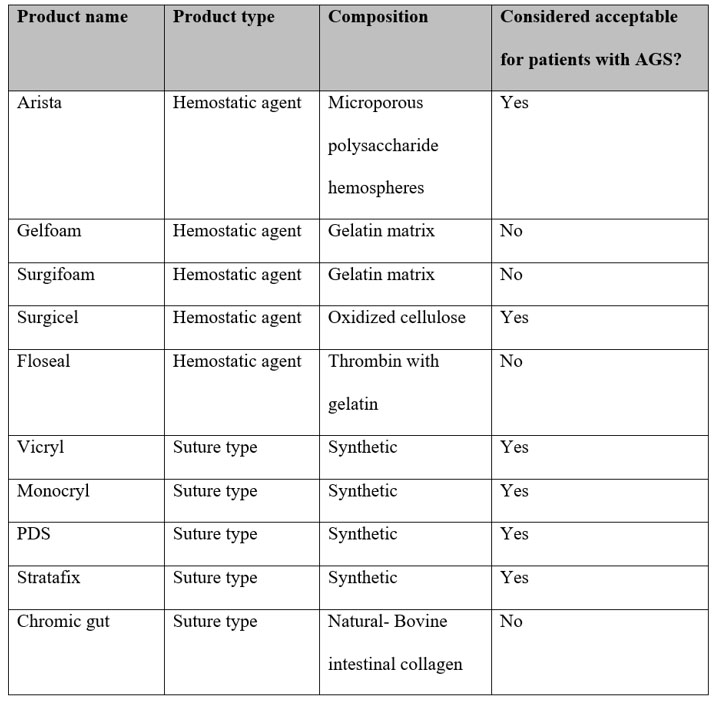
For patients with known or suspected AGS, preoperative planning should include a detailed allergy history and review of prior reactions to mammalian-derived foods, medications, and medical products. When the diagnosis is uncertain or the anticipated procedure may require mammalian-derived materials, allergist consultation and alpha-gal-specific IgE testing may assist with risk assessment. Importantly, perioperative exposure extends beyond medications to include hemostatic agents, graft materials, dural substitutes, biologic implants, and other products that may contain bovine- or porcine-derived components. Razzaq et al. found that 64% of 25 commonly used agents evaluated for spine surgery were potentially unsafe for patients with AGS, highlighting the difficulty of identifying these exposures based on product type alone [25]. The authors recommend pharmacy verification of product ingredients and substitution with nonmammalian alternatives when available, including plant-based polysaccharide powders, oxidized cellulose, human recombinant thrombin, and synthetic sealants [25]. Rinehart et al. similarly emphasize the perioperative implications of mammalian-derived medications and biologic materials, particularly in procedures where exposure to porcine- or bovine-derived products may be unavoidable [26].
Although the available literature consists primarily of case reports and expert recommendations, these reports consistently demonstrate that perioperative exposure to mammalian-derived products represents a clinically significant but often underrecognized source of risk for patients with AGS.
Management should therefore involve coordination among the surgical, anesthesia, pharmacy, and allergy teams before the procedure. Potentially high-risk medications and surgical materials should be identified in advance with alternatives selected when feasible (Figure 7). Real-time communication regarding AGS and anticipated exposures is particularly important because reactions to parenteral or implanted mammalian-derived products may occur during the procedure and can be severe. Henderson et al. reported profound intraoperative hemodynamic collapse in a patient with previously undiagnosed AGS undergoing cardiac surgery, illustrating the potential consequences when the diagnosis is not recognized before exposure [27]. For patients with established AGS, documenting the diagnosis prominently in the medical record and developing a procedure-specific plan for product selection may reduce inadvertent re-exposure. When mammalian-derived products cannot be avoided, individualized risk assessment and consideration of premedication should occur in consultation with allergy and anesthesia specialists [25],[26]. These precautions are especially relevant following a suspected perioperative reaction, as future procedures provide another opportunity for exposure to the same or similar mammalian-derived materials.
CONCLUSION
Perioperative awareness of AGS is paramount. While current literature primarily describes immediate hypersensitivity reactions to mammalian-derived surgical materials and delayed reactions following ingestion of mammalian products, this case demonstrates that intraoperatively applied mammalian-derived agents may contribute to delayed and prolonged inflammatory responses in sensitized patients. Although a causal relationship cannot be established from this case alone, the temporal course, persistent eosinophilia, recurrent postoperative complications, and prolonged presence of the bovine-derived hemostatic agent support consideration of an immune-mediated component.
This case identifies a gap in the medical literature regarding delayed postoperative reactions to mammalian-derived surgical materials in patients with AGS. Perioperative identification of AGS, careful review of medications and surgical materials, and multidisciplinary planning may help minimize inadvertent exposure and reduce the risk of hypersensitivity reactions. Following a suspected perioperative reaction, consideration of potential mammalian-derived exposures may also be important when evaluating otherwise unexplained or recurrent postoperative complications.
As this syndrome continues to rise in prevalence along with potential for significant patient morbidity and healthcare resource utilization, this topic warrants dedicated investigation in future studies to better define evidence-based strategies for perioperative risk reduction.
REFERENCES
1.
Centers for Disease Control and Prevention. About alpha-gal syndrome. Atlanta (GA): CDC; 2026. [Available at: https://www.cdc.gov/alpha-gal-syndrome/about/index.html]

2.
Wilson JM, Erickson L, Levin M, Ailsworth SM, Commins SP, Platts-Mills TAE. Tick bites, IgE to galactose-alpha-1,3-galactose and urticarial or anaphylactic reactions to mammalian meat: The alpha-gal syndrome. Allergy. 2024;79(6):1440–54. [CrossRef]
[Pubmed]
3.
Charles N, Blank U. IgE-mediated activation of mast cells and basophils in health and disease. Immunol Rev 2025;331(1):e70024. [CrossRef]
[Pubmed]
4.
Mollah F, Zacharek MA, Benjamin MR. What is alphagal syndrome? JAMA 2024;331(1):86. [CrossRef]
[Pubmed]
5.
Shishido AA, Wormser GP. A review of alpha-gal syndrome for the infectious diseases practitioner. Open Forum Infect Dis 2025;12(8):ofaf430. [CrossRef]
[Pubmed]
6.
Thompson JM, Carpenter A, Kersh GJ, Wachs T, Commins SP, Salzer JS. Geographic distribution of suspected alpha-gal syndrome cases—United States, January 2017–December 2022. MMWR Morb Mortal Wkly Rep 2023;72(30):815–20. [CrossRef]
[Pubmed]
7.
Binder AM, Commins SP, Altrich ML, Wachs T, Biggerstaff BJ, Beard CB, et al. Diagnostic testing for galactose-alpha-1,3-galactose, United States, 2010 to 2018. Ann Allergy Asthma Immunol 2021;126(4):411–6.e1. [CrossRef]
[Pubmed]
8.
Ross M, Daley G, Burnard ML, Sen A, Beyene E, Bisrat M, et al. Alpha-gal on the rise: The alarming growth of alpha-gal syndrome in high-risk regions. Cureus 2025;17(7):e88415. [CrossRef]
[Pubmed]
9.
Carpenter A, Drexler NA, McCormick DW, Thompson JM, Kersh G, Commins SP, et al. Health care provider knowledge regarding alpha-gal syndrome—United States, March–May 2022. MMWR Morb Mortal Wkly Rep 2023;72(30):809–14. [CrossRef]
[Pubmed]
10.
McGill SK, Hashash JG, Platts-Mills TA. AGA clinical practice update on alpha-gal syndrome for the GI clinician: Commentary. Clin Gastroenterol Hepatol 2023;21(4):891–6. [CrossRef]
[Pubmed]
11.
Kepley CL, Wang Y, Yelton A, Siebert ER, Iweala OI. Ticked off: Allergic effector cells in the pathogenesis of alpha-gal syndrome. Curr Allergy Asthma Rep 2025;25(1):57. [CrossRef]
[Pubmed]
12.
Platts-Mills TAE, Gangwar RS, Workman L, Wilson JM. The immunology of alpha-gal syndrome: History, tick bites, IgE, and delayed anaphylaxis to mammalian meat. Immunol Rev 2025;332(1):e70035. [CrossRef]
[Pubmed]
13.
Marwa K, Goldin J, Kondamudi NP. Type IV hypersensitivity reaction. In: StatPearls. Treasure Island (FL): StatPearls Publishing; 2025.
[Pubmed]
14.
Kim KM, D’Elia AM, Rodell CB. Hydrogel-based approaches to target hypersensitivity mechanisms underlying autoimmune disease. Adv Drug Deliv Rev 2024;212:115395. [CrossRef]
[Pubmed]
15.
Liu AW, Gillis JE, Sumpter TL, Kaplan DH. Neuroimmune interactions in atopic and allergic contact dermatitis. J Allergy Clin Immunol 2023;151(5):1169–77. [CrossRef]
[Pubmed]
16.
Baxter Healthcare Pty Ltd. FLOSEAL Hemostatic Matrix patient information leaflet. Baxter Healthcare; 2021. [Available at: https://www.baxterhealthcare.com.au/sites/g/files/ebysai1316/files/2021-11/FLOSEAL%20Patient%20Information%20 Leaflet_0.pdf]
17.
Baxter Healthcare Pty Ltd. FLOSEAL Hemostatic Matrix patient information leaflet. Baxter Healthcare; 2021. [Available at: https://www.baxterhealthcare.com.au/sites/g/files/ebysai1316/files/2021-11/FLOSEAL%20Patient%20Information%20Leaflet_0.pdf] [CrossRef]
[Pubmed]
18.
Carlstrom LP, Akhter A, Macielak RJ, Callejas CA, Eickstaedt JE, Brooks C, et al. Neurosurgical management of patients with alpha-gal syndrome. J Neurol Surg Rep 2024;85(3):e96–100. [CrossRef]
[Pubmed]
19.
Nourian MM, Stone CA Jr, Siegrist KK, Riess ML. Perioperative implications of patients with alpha gal allergies. J Clin Anesth 2023;86:111056. [CrossRef]
[Pubmed]
20.
Barkyoumb D, Tavakol SA, Zhao X, Stephens ME, Bageac DV, Bowen IE, et al. Povidone-iodine-induced scalp lesions in pediatric neurosurgery patients: A case series. Childs Nerv Syst 2024;41(1):27. [CrossRef]
[Pubmed]
21.
Kennedy H. Contact dermatitis secondary to povidone-iodine: A systematic review. Contact Dermatitis 2025;92(1):2–8. [CrossRef]
[Pubmed]
22.
Dalai R, Vinay S, Sai NSHC, Vineet J, Ramji M, Sayan B, et al. Experimental investigation of tensile properties and fracture behavior of ophthalmic sutures under straight, knotted, and looped configurations: Vicryl, nylon, and polypropylene. PLoS One 2026;21(2):e0343682. [CrossRef]
[Pubmed]
23.
Nalin F, Scarmozzino R, Arcolaci A, Olivieri B, Tommasi M, Bonadonna P, et al. Alpha-gal syndrome: When treatment of hypovolemic shock can lead to anaphylaxis. Allergol Immunopathol (Madr) 2024;52(3):60–4. [CrossRef]
[Pubmed]
24.
Lied GA, Lund KB, Storaas T. Intraoperative anaphylaxis to gelatin-based hemostatic agents: A case report. J Asthma Allergy 2019;12:163–7. [CrossRef]
[Pubmed]
25.
Razzaq BW, Passman JN, DiMaria S, Servider J, Kleyner RS, Sacks GI, et al. Complex spine surgery in the setting of alpha-gal syndrome. World Neurosurg 2026;205:124634. [CrossRef]
[Pubmed]
26.
Rinehart CD, Wang K, LoBue C, Augoustides JG, Patel SJ, Monette P, et al. Perioperative considerations for a patient with alpha-gal syndrome undergoing cardiac surgery. J Cardiothorac Vasc Anesth 2025;39(9):2516–23. [CrossRef]
[Pubmed]
27.
Henderson JJ, Saleem D, George E, Still S. Intraoperative anaphylaxis in the setting of undiagnosed alpha-gal syndrome: A heart team clinical conundrum. JACC Case Rep 2026;31(1):106033. [CrossRef]
[Pubmed]
SUPPORTING INFORMATION
Acknowledgments
Thank you, Dr. Sharon Smallwood, MD, for your assistance in gaining approval from Cherokee Nation Health Services to initiate this case report.
Author ContributionsBridget Botsford - Conception of the work, Design of the work, Acquisition of data, Analysis of data, Drafting the work, Revising the work critically for important intellectual content, Final approval of the version to be published, Agree to be accountable for all aspects of the work in ensuring that questions related to the accuracy or integrity of any part of the work are appropriately investigated and resolved.
Tschantre Dorsett - Conception of the work, Design of the work, Acquisition of data, Analysis of data, Revising the work critically for important intellectual content, Final approval of the version to be published, Agree to be accountable for all aspects of the work in ensuring that questions related to the accuracy or integrity of any part of the work are appropriately investigated and resolved.
Natasha Bray - Drafting the work, Revising the work critically for important intellectual content, Final approval of the version to be published, Agree to be accountable for all aspects of the work in ensuring that questions related to the accuracy or integrity of any part of the work are appropriately investigated and resolved.
Allisyn Shed - Drafting the work, Final approval of the version to be published, Agree to be accountable for all aspects of the work in ensuring that questions related to the accuracy or integrity of any part of the work are appropriately investigated and resolved.
Alexis Jones - Acquisition of data, Revising the work critically for important intellectual content, Final approval of the version to be published, Agree to be accountable for all aspects of the work in ensuring that questions related to the accuracy or integrity of any part of the work are appropriately investigated and resolved.
Guaranter of SubmissionThe corresponding author is the guarantor of submission.
Source of SupportNone
Consent StatementWritten informed consent was obtained from the patient for publication of this article.
Data AvailabilityAll relevant data are within the paper and its Supporting Information files.
Conflict of InterestAuthors declare no conflict of interest.
Copyright© 2026 Bridget Botsford et al. This article is distributed under the terms of Creative Commons Attribution License which permits unrestricted use, distribution and reproduction in any medium provided the original author(s) and original publisher are properly credited. Please see the copyright policy on the journal website for more information.