|
Case Report
Intrauterine device with double trouble: Bilateral tubal ectopic pregnancy in a woman with a copper intrauterine device
1 Physician Assistant, Emergency Department, Queens Hospital Center, New York City, United States; Chang Family Medical Center, Brooklyn, New York, United States
2 DO, Emergency Department, Queens Hospital Center, New York City, United States
Address correspondence to:
Kyaw Zaw Lin
Physician Assistant, Emergency Department, Queens Hospital Center, New York City, United States; Chang Family Medical Center, Brooklyn, New York,
United States
Message to Corresponding Author
Article ID: 100046G06KL2026
Access full text article on other devices

Access PDF of article on other devices


How to cite this article
Lin KZ, Kernizan S. Intrauterine device with double trouble: Bilateral tubal ectopic pregnancy in a woman with a copper intrauterine device. Edorium J Gynecol Obstet 2026;10(2):8–14.ABSTRACT
Introduction: Ectopic pregnancy refers to implantation of a gestation outside the endometrial cavity and remains a major contributor to early pregnancy morbidity. While ectopic pregnancies are typically unilateral, bilateral tubal implantation represents an uncommon clinical entity with an estimated incidence of approximately 1 per 200,000 pregnancies. Pregnancy occurring with an intrauterine device (IUD) in situ is uncommon; however, when conception occurs, the relative risk of ectopic implantation is increased. We report a rare case of bilateral tubal ectopic pregnancy associated with a copper IUD that was successfully managed with medical therapy.
Case Report: A 26-year-old nulligravid woman with a copper IUD in place since 2020 presented after two positive home pregnancy tests. She was asymptomatic, denying abdominal pain, vaginal bleeding, dizziness, or urinary complaints. Vital signs were stable, and laboratory studies were normal except for a serum β-human chorionic gonadotropin (β-hCG) level of 4655 mIU/mL. Transabdominal and transvaginal ultrasonography demonstrated a normal uterus without gross anatomical or structural defect and a correctly positioned IUD without an intrauterine gestational sac. Bilateral complex adnexal masses measuring 2.4 × 1.6 × 1.4 cm on the right and 2.9 × 1.7 × 2.2 cm on the left, each demonstrating peripheral vascularity, were highly suspicious for bilateral ectopic pregnancy. No significant pelvic free fluid was seen. The IUD was removed, and intramuscular methotrexate (MTX) (50 mg/m2 ; 88.5 mg) was administered. Because the β-hCG level increased to 4924 mIU/mL on day 4, a second MTX dose was given. Serial β-hCG measurements subsequently declined appropriately, reaching 201 mIU/mL by day 17, confirming successful resolution without surgery or complications.
Conclusion: The uniqueness of this case lies in the occurrence of bilateral tubal ectopic pregnancy in an asymptomatic patient with a copper IUD who achieved successful medical resolution. Early recognition through meticulous ultrasonographic evaluation of both adnexa, combined with serial β-hCG monitoring and appropriate patient selection, permitted successful fertility-preserving medical management while avoiding surgery and its associated risks.
Keywords: Bilateral ectopic pregnancy, Copper intrauterine device, Medical management, Methotrexate
INTRODUCTION
Intrauterine devices (IUDs) are widely recognized as one of the most effective long-acting reversible contraceptive methods because of their high contraceptive efficacy, established safety, and excellent tolerability. Despite their favorable safety profile, complications may occur during insertion or throughout the duration of use. Common complications include device expulsion (3–6% within the first year), pelvic inflammatory disease (approximately 1% within the first 20 days after insertion and 0.5% during the following 3–6 months), contraceptive failure (0.1–0.6% during the first year), uterine perforation (approximately 0.01%), and device malposition, which may occur in up to 10% of users but is often clinically insignificant. Although pregnancy with an IUD is uncommon, it carries an increased risk of ectopic implantation. Pelvic pain and abnormal uterine bleeding are the most common adverse effects and account for many early device removals. Early recognition and timely management of these complications are critical for optimizing patient safety, improving patient satisfaction, and promoting continued use of effective contraception [1],[2].
CASE REPORT
A 26-year-old nulligravid woman with a copper intrauterine device (IUD) placed in 2020 presented to the emergency department after obtaining two positive home pregnancy tests four days earlier. She sought medical evaluation to confirm the pregnancy and was completely asymptomatic at presentation. She denied vaginal bleeding, spotting, pelvic or abdominal pain, dysuria, hematuria, flank pain, fever, chills, nausea, vomiting, diarrhea, constipation, dizziness, chest pain, dyspnea, weakness, or neurologic symptoms. She had no history of prior pregnancy, ectopic pregnancy, pelvic inflammatory disease, infertility, or previous pelvic surgery. The patient was hemodynamically stable with normal vital signs. Physical examination demonstrated a soft, non-distended abdomen without tenderness, guarding, rebound tenderness, or adnexal tenderness.
Laboratory evaluation revealed a white blood cell count of 9.11 × 10³/µL without a left shift, hemoglobin of 12.4 g/dL, hematocrit of 36.9%, mean corpuscular volume of 97.4 FL, and platelet count of 310 × 103/µL, with no evidence of anemia or active hemorrhage. Renal and hepatic function were normal (blood urea nitrogen, 12 mg/dL; creatinine, 0.69 mg/dL; estimated glomerular filtration rate, 123 mL/min/1.73 m2; aspartate aminotransferase, 16 U/L; alanine aminotransferase, 6 U/L; alkaline phosphatase, 92 U/L), supporting eligibility for MTX therapy if indicated.
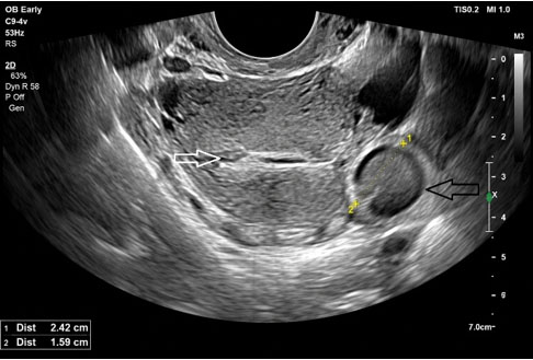
Given the positive pregnancy test with an intrauterine device in situ, pelvic ultrasonography was performed to determine the location and viability of the pregnancy. Transabdominal ultrasonography demonstrated a normal-sized uterus (9.1 × 5.2 × 5.3 cm) with the copper intrauterine device appropriately positioned within the endometrial cavity. An intrauterine gestational sac was not identified (Figure 1). The uterus was otherwise unremarkable, without evidence of fibroids or congenital anomalies, and no significant pelvic free fluid was observed.
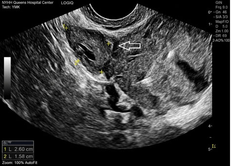
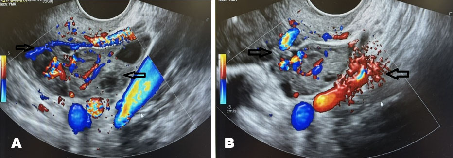
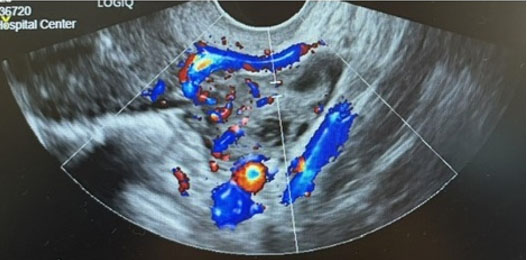
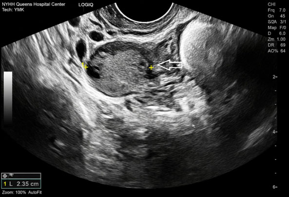
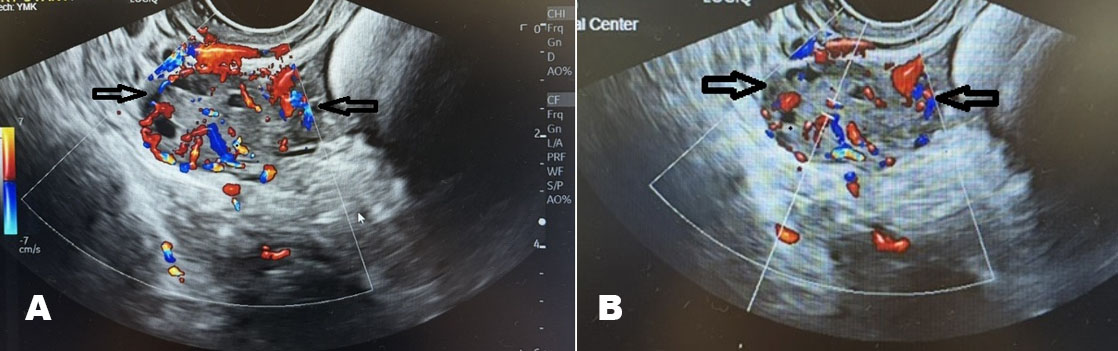
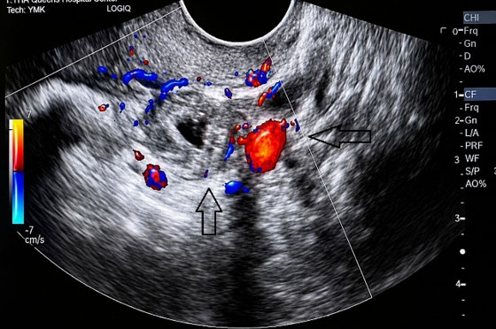
Further evaluation with transvaginal ultrasonography confirmed the absence of an intrauterine pregnancy despite the elevated β-human chorionic gonadotropin (β-hCG) level. Examination of the left adnexa identified a complex left adnexal mass measuring 2.9 × 1.7 × 2.2 cm, which demonstrated peripheral vascular flow on color Doppler imaging, a finding highly suggestive of left ectopic pregnancy (Figure 2, Figure 3, Figure 4). A small hemorrhagic follicle measuring 1.6 × 1.4 × 1.1 cm was also noted within the left ovary. Importantly, no significant pelvic free fluid or sonographic evidence of tubal rupture or hemoperitoneum was present. A second complex right adnexal mass measuring 2.4 × 1.6 × 1.4 cm had similar sonographic characteristics, a finding highly suggestive of right ectopic pregnancy (Figure 5, Figure 6, Figure 7), raising strong suspicion for bilateral tubal ectopic pregnancy.
The combination of a positive pregnancy test, absence of an intrauterine gestational sac, bilateral vascular adnexal masses, and a correctly positioned IUD established the diagnosis of bilateral tubal ectopic pregnancy. Although bilateral ectopic pregnancy is exceedingly uncommon, the characteristic ultrasonographic findings involving both adnexa allowed diagnosis before rupture or surgical exploration.
Because the patient remained asymptomatic and hemodynamically stable without evidence of rupture, conservative medical management was considered appropriate. The copper IUD was removed under sterile conditions, and systemic MTX was administered using the standard single-dose protocol at 50 mg/m2 (total dose, 88.5 mg intramuscularly). The patient received comprehensive counseling regarding treatment expectations, potential adverse effects of MTX, signs and symptoms of tubal rupture, and the importance of strict follow-up with serial serum β-hCG concentration.
Four days after the initial MTX dose, the serum β-hCG level increased slightly to 4924 mIU/mL, an expected early post-treatment response that did not meet criteria for an adequate therapeutic decline. Accordingly, a second intramuscular dose of MTX (88.5 mg) was administered per established treatment protocols. Serial β-hCG measurements subsequently demonstrated an appropriate biochemical response, declining to 3929 mIU/mL on day 7 (>15% decrease from day 4), 2421 mIU/mL on day 10, 1206 mIU/mL on day 14, and 201 mIU/mL on day 17. These findings confirmed successful medical resolution of the bilateral ectopic pregnancies without surgical intervention. Throughout treatment, the patient remained clinically stable without abdominal pain, vaginal bleeding, hemodynamic instability, or evidence of tubal rupture. She experienced no adverse effects related to MTX therapy and required neither hospitalization nor emergency surgical intervention. The favorable clinical course illustrates that carefully selected patients with bilateral tubal ectopic pregnancy can be successfully managed with systemic MTX when diagnosed early, remain hemodynamically stable, and adhere to close biochemical surveillance.
DISCUSSION
Although intrauterine devices are among the most effective forms of reversible contraception and significantly reduce the overall risk of pregnancy, conception occurring with an IUD in situ carries a disproportionately increased likelihood of ectopic implantation. Reported rates of ectopic pregnancy among women who conceive while using a copper IUD range from approximately 11% to 15%, whereas rates among users of the 52-mg levonorgestrel-releasing intrauterine device (LNG-IUD) range from 27% to 53%. Large population-based studies further demonstrate that the absolute incidence of ectopic pregnancy varies according to the type of LNG-IUD, emphasizing that any pregnancy diagnosed in a patient with an IUD requires prompt evaluation to exclude an ectopic gestation [3],[4].
Pregnancy in the presence of an IUD is rare but may result from several factors affecting contraceptive efficacy. Expulsion, whether complete or partial and particularly when unrecognized, can compromise contraceptive protection. Similarly, malposition or suboptimal placement, including displacement from the uterine fundus toward the lower uterine segment or cervical canal, may diminish contraceptive effectiveness. Although uncommon, uterine perforation with extrauterine migration of the device represents another potential mechanism of contraceptive failure [5]. Nevertheless, conception can occur even when the IUD remains appropriately positioned within the uterine cavity, reflecting the small inherent failure rate of these highly effective contraceptive methods [6]. Copper IUDs exert their contraceptive effect predominantly through local intrauterine reactions that adversely affect sperm viability, motility, and function, thereby interfering with fertilization; alterations of the endometrial environment may provide an additional contraceptive effect [7]. Therefore, when pregnancy occurs despite confirmation of appropriate IUD positioning, the event may represent an uncommon failure of the device’s contraceptive mechanisms rather than expulsion or malposition as in our case report.
Ectopic pregnancy, an implantation of a fertilized ovum outside the uterine cavity, accounts for approximately 1% to 2% of all pregnancies. Tubal implantation accounts for the vast majority of ectopic pregnancies, with the ampullary region representing the predominant site, although implantation may also occur in the interstitial portion of the tube, cervix, ovary, cesarean scar, myometrium, or abdominal cavity [8]. Bilateral tubal ectopic pregnancy is exceptionally uncommon, making diagnosis particularly challenging because clinical manifestations often resemble those of unilateral disease. Consequently, careful evaluation of both adnexa during transvaginal ultrasonography is essential, especially in pregnancies occurring with an IUD. The incidence of ectopic pregnancy varies among different populations and has changed over time because of improvements in diagnostic imaging and early pregnancy surveillance. Population-based studies have reported incidences ranging from approximately 6 to 21 ectopic pregnancies per 1000 pregnancies [9],[10]. Despite advances in transvaginal ultrasonography and quantitative serum β-hCG testing that have improved early diagnosis, ectopic pregnancy remains a major cause of maternal morbidity and first-trimester pregnancy-related mortality. Significant racial disparities persist, with Black women experiencing disproportionately higher rates of ectopic pregnancy-related morbidity and mortality than White women, underscoring ongoing inequities in healthcare access, timely diagnosis, and clinical outcomes [11],[12],[13].
Several established risk factors increase the likelihood of ectopic pregnancy. Previous ectopic pregnancy is the strongest predictor, conferring a seven- to eleven-fold increased risk of recurrence. Additional risk factors for ectopic pregnancy include prior pelvic or abdominal surgery, pelvic inflammatory disease, infertility, cigarette smoking, induced abortion, previous ectopic pregnancy, and underlying tubal pathology. These factors disrupt normal tubal anatomy or function, impair embryo transport, and increase the likelihood of extrauterine implantation. Prior salpingostomy is associated with an elevated risk of recurrent ectopic pregnancy and warrants heightened clinical vigilance in subsequent pregnancies [14],[15].
Surgical management used to be the standard treatment for ectopic pregnancy. However, improvements in early diagnosis have expanded the role of conservative management. In carefully selected hemodynamically stable patients without evidence of tubal rupture, MTX has become the preferred first-line treatment and achieves overall success rates approaching 90% [16]. Early diagnosis facilitates medical management, decreases the need for surgery, preserves reproductive potential, and reduces treatment-related morbidity. In selected patients with low and spontaneously declining β-hCG concentrations, expectant management may also be appropriate, provided close clinical and biochemical surveillance can be maintained [17],[18].
Appropriate patient selection is critical for successful MTX therapy. Candidates for MTX therapy should be hemodynamically stable, have no evidence of tubal rupture or fetal cardiac activity, and be able to comply with serial follow-up and access emergency care if needed. A serum β-hCG concentration of approximately 5000 mIU/mL or less is commonly used as a practical threshold for medical management, although successful treatment has been reported at higher levels in selected patients. Most treatment protocols also recommend an ectopic mass measuring less than 3 to 4 cm; however, current evidence suggests that lesion size alone is a less reliable predictor of treatment success than baseline β-hCG concentration [19],[20],[21],[22],[23].
Patients with rupture, hemodynamic compromise, or significant organ dysfunction require surgical rather than medical management with MTX. In these situations, surgical management remains the preferred treatment because delayed intervention may result in catastrophic hemorrhage or life-threatening complications. Among the predictors of MTX failure, elevated baseline β-hCG concentration remains the most important. Patients with β-hCG levels greater than 5000 mIU/mL are significantly more likely to require additional MTX doses or subsequent surgical intervention than those with lower hormone concentrations. Similarly, the presence of embryonic cardiac activity on transvaginal ultrasonography is strongly associated with treatment failure and is generally considered a relative contraindication to medical management. Although larger ectopic masses have traditionally been associated with reduced success, evidence supporting mass size as an independent predictor remains inconsistent [20],[21]. Tubal rupture remains the most serious complication of ectopic pregnancy and is responsible for the majority of ectopic pregnancy-related deaths. The reported mortality rate is approximately 31.9 per 100,000 pregnancies [24].
Concerns regarding the adverse effects of MTX on future fertility have not been supported by available evidence. Studies indicate that MTX does not negatively impact ovarian reserve or future reproductive outcomes, including assisted reproductive techniques, supporting its role as a safe and fertility-preserving treatment option in appropriately selected patients [25],[26].
The commonly used regimen is a single intramuscular dose of MTX (50 mg/m2), with serial β-hCG monitoring on treatment days 1, 4, and 7. A transient increase in β-hCG between days 1 and 4 is expected due to ongoing syncytiotrophoblastic hormone production despite inhibition of cytotrophoblast proliferation. Successful treatment is indicated by a decline of at least 15% between days 4 and 7, after which weekly β-hCG monitoring is continued until complete biochemical resolution [27]. Approximately 15% to 20% of patients require a second MTX dose because of an inadequate decline in β-hCG levels, whereas fewer than 1% require more than two doses. Our patient demonstrated the expected transient rise in β-hCG after the initial dose and subsequently responded appropriately following administration of a second dose, achieving complete biochemical resolution without surgery [28].
Routine serial ultrasonography following MTX treatment is generally unnecessary because enlargement of the ectopic mass frequently reflects hematoma formation rather than persistent trophoblastic tissue. Repeat imaging should be reserved for patients who develop worsening abdominal pain or clinical findings concerning for tubal rupture. Downtrending in serum β-hCG levels does not exclude the risk of tubal rupture; therefore, clinicians should prompt consideration in patients who develop severe abdominal pain or hemodynamic instability. Patients treated with MTX require comprehensive counseling regarding treatment expectations and strict follow-up. Patients are advised to refrain from becoming pregnant until serum β-hCG levels have returned to undetectable values. Additionally, they should not take folic acid supplements, limit prolonged sunlight exposure, and, when possible, discontinue medications known to affect MTX metabolism, such as nonsteroidal anti-inflammatory drugs. Mild abdominal pain occurring approximately one week after treatment is relatively common and is usually self-limited; however, severe or progressive pain requires urgent evaluation to exclude rupture or significant hemoperitoneum [29].
Surgical intervention remains the treatment of choice for patients with tubal rupture, hemodynamic instability, contraindications to MTX, or persistent disease despite medical therapy. The principal operative procedures are salpingectomy and salpingostomy. Salpingectomy completely removes the affected fallopian tube, whereas salpingostomy preserves tubal anatomy through removal of the ectopic gestation alone. Although salpingostomy offers the advantage of fertility preservation, it carries a greater risk of persistent trophoblastic tissue and recurrent ectopic pregnancy. Nevertheless, long-term fertility outcomes appear comparable between both procedures when patients are appropriately selected [30],[31].
The present case illustrates several important clinical lessons. First, pregnancy occurring in the presence of an IUD should always prompt immediate evaluation for ectopic pregnancy. Second, bilateral adnexal assessment is essential because bilateral tubal ectopic pregnancy, although exceedingly rare, may be overlooked if only one adnexa is examined. Finally, early diagnosis combined with careful patient selection and close β-hCG surveillance can facilitate successful nonsurgical management, preserve reproductive potential, and prevent the significant morbidity and mortality associated with delayed diagnosis or tubal rupture.
CONCLUSION
Bilateral tubal ectopic pregnancy associated with an intrauterine device is an exceptionally rare but potentially life-threatening condition that requires a high index of clinical suspicion, even in asymptomatic patients. This case highlights the importance of prompt pregnancy evaluation with transvaginal ultrasonography and careful assessment of both adnexa whenever pregnancy occurs in the presence of an IUD. Early detection before tubal rupture allows consideration of fertility-preserving treatment options and reduces the risk of significant maternal morbidity.
In appropriately selected patients, systemic MTX provides an effective nonsurgical alternative to operative management. Treatment success with MTX is greatest when the initial serum β hCG level is ≤5000 mIU/mL; however, carefully selected patients with higher β-hCG concentrations may also achieve successful medical management. Serial serum β-hCG monitoring is critical for evaluating therapeutic response and identifying patients who require additional MTX dosing or surgical intervention.
In this patient, early identification of bilateral adnexal pathology before rupture allowed successful medical treatment and avoided operative intervention while preserving future fertility. Clinicians require careful evaluation for bilateral ectopic pregnancy in patients who conceive with an intrauterine device in situ, irrespective of symptom severity.
REFERENCES
1.
Braaten KP, Benson CB, Maurer R, Goldberg AB. Malpositioned intrauterine contraceptive devices: Risk factors, outcomes, and future pregnancies. Obstet Gynecol 2011;118(5):1014–20. [CrossRef]
[Pubmed]

2.
Grunloh DS, Casner T, Secura GM, Peipert JF, Madden T. Characteristics associated with discontinuation of long-acting reversible contraception within the first 6 months of use. Obstet Gynecol 2013;122(6):1214–21. [CrossRef]
[Pubmed]
3.
Xiong X, Buekens P, Wollast E. IUD use and the risk of ectopic pregnancy: A meta-analysis of case-control studies. Contraception 1995;52(1):23–34. [CrossRef]
[Pubmed]
4.
Meaidi A, Torp-Pedersen C, Lidegaard Ø, Mørch LS. Ectopic pregnancy risk in users of levonorgestrel-releasing intrauterine systems with 52, 19.5, and 13.5 mg of hormone. JAMA 2023;329(11):935–7. [CrossRef]
[Pubmed]
5.
Boortz HE, Margolis DJA, Ragavendra N, Patel MK, Kadell BM. Migration of intrauterine devices: Radiologic findings and implications for patient care. Radiographics 2012;32(2):335–52. [CrossRef]
[Pubmed]
6.
Thonneau P, Goulard H, Goyaux N. Risk factors for intrauterine device failure: A review. Contraception 2001;64(1):33–7. [CrossRef]
[Pubmed]
7.
Stanford JB, Mikolajczyk RT. Mechanisms of action of intrauterine devices: Update and estimation of postfertilization effects. Am J Obstet Gynecol 2002;187(6):1699–708. [CrossRef]
[Pubmed]
8.
Bouyer J, Coste J, Fernandez H, Pouly JL, JobSpira N. Sites of ectopic pregnancy: A 10 year population-based study of 1800 cases. Hum Reprod 2002;17(12):3224–30. [CrossRef]
[Pubmed]
9.
Van Den Eeden SK, Shan J, Bruce C, Glasser M. Ectopic pregnancy rate and treatment utilization in a large managed care organization. Obstet Gynecol 2005;105(5 Pt 1):1052–7. [CrossRef]
[Pubmed]
10.
Hoover KW, Tao G, Kent CK. Trends in the diagnosis and treatment of ectopic pregnancy in the United States. Obstet Gynecol 2010;115(3):495–502. [CrossRef]
[Pubmed]
11.
Stulberg DB, Cain LR, Dahlquist I, Lauderdale DS. Ectopic pregnancy rates and racial disparities in the Medicaid population, 2004–2008. Fertil Steril 2014;102(6):1671–6. [CrossRef]
[Pubmed]
12.
Mann LM, Kreisel K, Llata E, Hong J, Torrone EA. Trends in ectopic pregnancy diagnoses in United States emergency departments, 2006–2013. Matern Child Health J 2020;24(2):213–21. [CrossRef]
[Pubmed]
13.
Stulberg DB, Cain L, Dahlquist IH, Lauderdale DS. Ectopic pregnancy morbidity and mortality in low-income women, 2004–2008. Hum Reprod 2016;31(3):666–71. [CrossRef]
[Pubmed]
14.
Brim ACS, Barretto VRD, Reis-Oliveira JG, da Silveira de Araújo RB, Romeo ACDCB. Risk factors for ectopic pregnancy occurrence: Systematic review and meta-analysis. Int J Gynaecol Obstet 2025;168(3):919–32. [CrossRef]
[Pubmed]
15.
Zhang D, Shi W, Li C, Yuan JJ, Xia W, Xue RH, et al. Risk factors for recurrent ectopic pregnancy: A case-control study. BJOG 2016;123(Suppl 3):82–9. [CrossRef]
[Pubmed]
16.
Lipscomb GH. Medical therapy for ectopic pregnancy. Semin Reprod Med 2007;25(2):93–8. [CrossRef]
[Pubmed]
17.
Barnhart KT, Gosman G, Ashby R, Sammel M. The medical management of ectopic pregnancy: A meta-analysis comparing “single dose” and “multidose” regimens. Obstet Gynecol 2003;101(4):778–84. [CrossRef]
[Pubmed]
18.
Morlock RJ, Lafata JE, Eisenstein D. Cost-effectiveness of single-dose methotrexate compared with laparoscopic treatment of ectopic pregnancy. Obstet Gynecol 2000;95(3):407–12. [CrossRef]
[Pubmed]
19.
Lipscomb GH, Meyer NL, Flynn DE, Peterson M, Ling FW. Oral methotrexate for treatment of ectopic pregnancy. Am J Obstet Gynecol 2002;186(6):1192–5. [CrossRef]
[Pubmed]
20.
Menon S, Colins J, Barnhart KT. Establishing a human chorionic gonadotropin cutoff to guide methotrexate treatment of ectopic pregnancy: A systematic review. Fertil Steril 2007;87(3):481–4. [CrossRef]
[Pubmed]
21.
Tsakiridis I, Giouleka S, Mamopoulos A, Athanasiadis A, Dagklis T. Diagnosis and management of ectopic pregnancy: A comparative review of major national guidelines. Obstet Gynecol Surv 2020;75(10):611–23. [CrossRef]
[Pubmed]
22.
Gamzu R, Almog B, Levin Y, Pauzner D, Lessing JB, Jaffa A, et al. The ultrasonographic appearance of tubal pregnancy in patients treated with methotrexate. Hum Reprod 2002;17(10):2585–7 [CrossRef]
[Pubmed]
23.
Grimes DA. Estimation of pregnancy-related mortality risk by pregnancy outcome, United States, 1991 to 1999. Am J Obstet Gynecol 2006;194(1):92–4. [CrossRef]
[Pubmed]
24.
Oriol B, Barrio A, Pacheco A, Serna J, Zuzuarregui JL, Garcia-Velasco JA. Systemic methotrexate to treat ectopic pregnancy does not affect ovarian reserve. Fertil Steril 2008;90(5):1579–82. [CrossRef]
[Pubmed]
25.
Wiser A, Gilbert A, Nahum R, Orvieto R, Haas J, Hourvitz A, et al. Effects of treatment of ectopic pregnancy with methotrexate or salpingectomy in the subsequent IVF cycle. Reprod Biomed Online 2013;26(5):449–53. [CrossRef]
[Pubmed]
26.
Gay C, Perrin J, Courbiere B, Bretelle F, Agostini A. Impact of salpingectomy for ectopic pregnancy on the ovarian response during IVF stimulation. J Gynecol Obstet Hum Reprod 2019;48(9):727–30. [CrossRef]
[Pubmed]
27.
American College of Obstetricians and Gynecologists’ Committee on Practice Bulletins—Gynecology. ACOG Practice Bulletin No. 193: Tubal Ectopic Pregnancy. Obstet Gynecol 2018;131(3):e91–103. [CrossRef]
[Pubmed]
28.
Natale A, Candiani M, Barbieri M, Calia C, Odorizzi MP, Busacca M. Pre- and post-treatment patterns of human chorionic gonadotropin for early detection of persistence after a single dose of methotrexate for ectopic pregnancy. Eur J Obstet Gynecol Reprod Biol 2004;117(1):87–92. [CrossRef]
[Pubmed]
29.
Verity L, Ludlow J, Dickinson JE. Interstitial ectopic pregnancy: A contemporary case series. Aust N Z J Obstet Gynaecol 2003;43(3):232–5. [CrossRef]
[Pubmed]
30.
Tang A, Baartz D, Khoo SK. A medical management of interstitial ectopic pregnancy: A 5-year clinical study. Aust N Z J Obstet Gynaecol 2006;46(2):107–11. [CrossRef]
[Pubmed]
31.
Lipscomb GH, Puckett KJ, Bran D, Ling FW. Management of separation pain after single-dose methotrexate therapy for ectopic pregnancy. Obstet Gynecol 1999;93(4):590–3. [CrossRef]
[Pubmed]
SUPPORTING INFORMATION
Author Contributions
Kyaw Zaw Lin - Conception of the work, Design of the work, Acquisition of data, Drafting the work, Revising the work critically for important intellectual content, Final approval of the version to be published, Agree to be accountable for all aspects of the work in ensuring that questions related to the accuracy or integrity of any part of the work are appropriately investigated and resolved.
Soraya Kernizan - Conception of the work, Design of the work, Analysis of data, Drafting the work, Revising the work critically for important intellectual content, Final approval of the version to be published, Agree to be accountable for all aspects of the work in ensuring that questions related to the accuracy or integrity of any part of the work are appropriately investigated and resolved.
Guaranter of SubmissionThe corresponding author is the guarantor of submission.
Source of SupportNone
Consent StatementWritten informed consent was obtained from the patient for publication of this article.
Data AvailabilityAll relevant data are within the paper and its Supporting Information files.
Conflict of InterestAuthors declare no conflict of interest.
Copyright© 2026 Kyaw Zaw Lin et al. This article is distributed under the terms of Creative Commons Attribution License which permits unrestricted use, distribution and reproduction in any medium provided the original author(s) and original publisher are properly credited. Please see the copyright policy on the journal website for more information.