|
Case Report
Vulvar melanoma: A rare case report from the department of obstetrics and gynecology
1 Resident in Obstetrics and Gynecology, CHU Mohammed VI, Agadir, Morocco
Address correspondence to:
Meriem Nouri
Resident in Obstetrics and Gynecology, CHU Mohammed VI, Agadir,
Morocco
Message to Corresponding Author
Article ID: 100044G06MN2026
Access full text article on other devices

Access PDF of article on other devices


How to cite this article
Nouri M, Baloul O, El Borgi H, Kouchih A, Ammouri S, El Farouqi A. Vulvar melanoma: A rare case report from the department of obstetrics and gynecology. Edorium J Gynecol Obstet 2026;11(1):1–8.ABSTRACT
Introduction: Approximately 3% to 5% of all cancer cases in women are caused by vulvar cancer, a rare tumor compared to other gynecological tumors. It is a particularly aggressive tumor originating from melanocytes and accounts for only 2.9% of all vulvar malignancies.
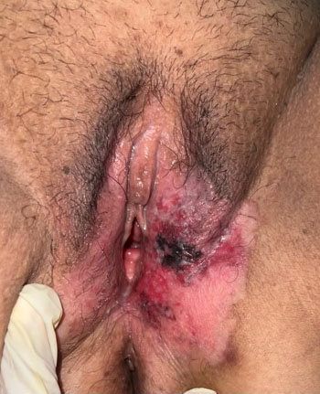
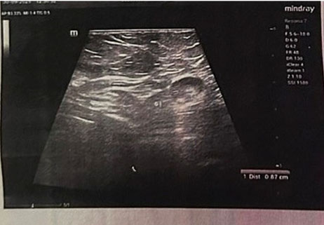
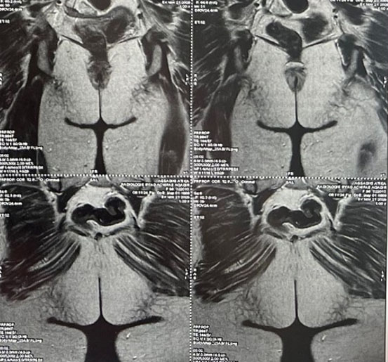
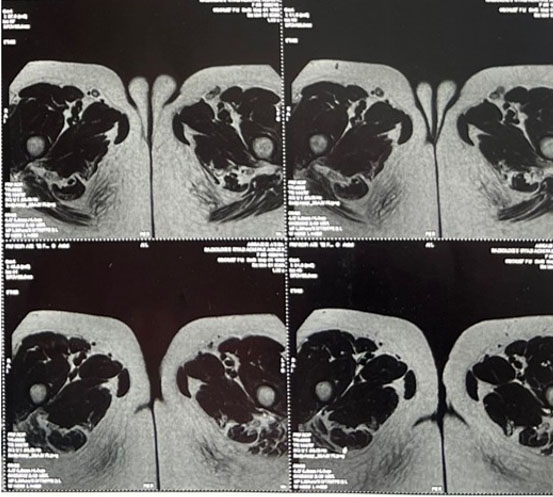
Case Report: A 56-year-old woman, single, menopausal for five years, presented with chronic left-sided vulvar pruritus evolving for approximately five years. Gynecological examination revealed a large erythematous lesion involving both labia majora, with a central erosive area and peripheral induration, associated with an irregular pigmented plaque centered on the left labium majus, extending posteriorly to the perineum and to the inner surface of the buttocks. Multiple excoriations due to scratching were noted, as well as several nevi, with no palpable inguinal lymphadenopathy. Bilateral inguinal ultrasound demonstrated lymphadenopathies of variable sizes, including nodes showing loss of fatty hilum, the largest measuring 10 × 9.4 mm on the left and 7 × 6 mm on the right, without abnormal fluid collection. Pelvic magnetic resonance imaging (MRI) revealed a vulvar tissue process located in the posterior aspect of the left labium majus, without signs of deep extension. Thoraco-abdomino-pelvic computed tomography (CT) showed no evidence of metastatic disease. The patient underwent partial vulvectomy combined with bilateral inguinal lymph node dissection followed by reconstruction using a flap procedure. Surgical margins were clear on frozen section. Final histopathological examination confirmed an invasive superficial spreading vulvar melanoma, associated with an extensive in situ component. Breslow thickness was 1 mm, with invasion limited to the superficial dermis (Chung level I). Mitotic activity was 6 mitoses per 10 high-power fields, with no ulceration, no lymphovascular invasion, and no perineural invasion. The invasive component was located more than 10 mm from all surgical margins; however, the in situ component involved the external and internal margins and was 6 mm from the superior margin. Bilateral inguinal lymph node dissection showed no metastatic involvement.
Conclusion: Vulvar melanoma is a rare and particularly aggressive tumor with a poor prognosis. Prognostic factors include tumor size, depth of invasion, tumor differentiation, surgical margins, patient age, involvement of adjacent structures, and especially lymph node status.
Keywords: Ainguinal lymphadenopathy, Case report, Vulvar melanoma
INTRODUCTION
Vulvar cancer is a rare tumor compared to other gynecological tumors and accounts for approximately 3% to 5% of all cancer cases in women [1]. It is estimated that 2 to 3 cases per 100,000 women occur each year, and it mainly affects women who are post-menopausal, typically between the ages of 50 and 70 [2]. The most prevalent cancer in men is squamous cell carcinoma, adenocarcinoma, basal cell carcinoma, malignant melanoma, sarcoma, and metastatic carcinomas. Among these tumors, vulvar melanoma (VM) ranks second after squamous cell carcinoma, representing 5% to 6% of all vulvar cancers [3]. Its occurrence seems to be increasing gradually [4]. It is a particularly aggressive tumor, originating from melanocytes, and accounts for only 2.9% of all vulvar malignancies. It most often occurs after menopause, usually around the sixth decade of life [1]. Clinically, its presentation is not very specific: bleeding and chronic itching are the most observed symptoms, with a wide variation in severity. Vulvar melanomas are rare tumors, often associated with a poor prognosis. On the molecular level, they are clearly different from cutaneous melanomas: while the latter commonly have a BRAF mutation, VMs are more often characterized by c-KIT mutations [5]. To confirm the melanocytic nature of the lesion, diagnosis requires a thorough histopathological examination. To treat, a multidisciplinary approach is necessary, which includes surgery, lymph node evaluation, and potentially targeted therapies or immunotherapies based on the molecular profile. The high risk of metastatic spread is the main reason why the prognosis remains poor despite these strategies [6].
CASE REPORT
A 56-year-old woman, single, nulligravid, and menopausal for five years, with a medical history of type 2 diabetes mellitus treated with oral antidiabetic agents, presented with a chronic left-sided vulvar pruritus evolving for approximately five years, refractory to medical treatment and without associated symptoms. Two years prior to consultation, the patient noticed the appearance of a darkly pigmented vulvar lesion, which prompted evaluation by a dermatologist. A vulvar biopsy was performed, revealing VM, after which the patient was referred to our department for further management.
On physical examination, the patient was in good general condition. Vital signs were stable, conjunctivae were normally colored.
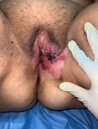
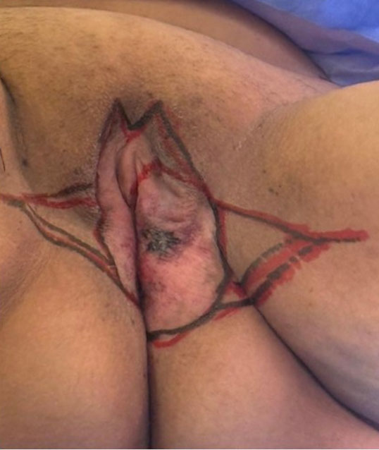
Gynecological examination revealed a large erythematous lesion involving both labia majora, with a central erosive area and peripheral induration, associated with an irregular pigmented plaque centered on the left labium majus, extending posteriorly to the perineum and to the inner surface of the buttocks. Multiple excoriations due to scratching were noted, as well as several nevi (Figure 1 and Figure 2). A speculum and bimanual examination were deferred due to the presence of an intact hymen. No palpable inguinal lymphadenopathy was detected. The remainder of the systemic examination was unremarkable.
Histopathological examination of two vulvar biopsy specimens revealed distinct findings. The first biopsy demonstrated a superficial spreading vulvar mucosal melanoma in situ, without any evidence of stromal invasion. The second biopsy showed an invasive superficial spreading vulvar mucosal melanoma, characterized by focal superficial stromal infiltration measuring approximately 1 mm, corresponding to Chung level I. The tumor thickness (Breslow depth) was 1 mm. No vascular emboli or perineural invasion were identified. An extensive pagetoid spread was also noted.
Bilateral inguinal ultrasound demonstrated bilateral lymphadenopathies of variable sizes, including nodes showing loss of fatty hilum. The largest nodes measured 10 × 9.4 mm on the left and 7 × 6 mm on the right without any abnormal fluid collection (Figure 3).
Pelvic magnetic resonance imaging (MRI) revealed a vulvar tissue process located in the posterior aspect of the left labium majus, without signs of deep extension (Figure 4 and Figure 5). Thoraco-abdomino-pelvic computed tomography (CT) showed no evidence of metastatic disease.
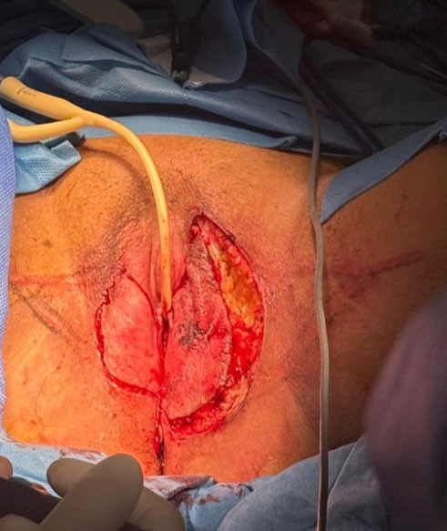
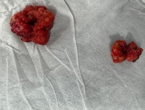
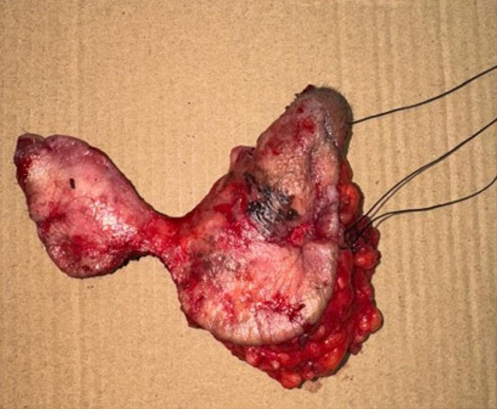
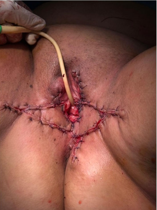
The patient underwent a partial vulvectomy combined with bilateral inguinal lymph node dissection (Figure 6, Figure 7, Figure 8, Figure 9), followed by reconstruction using a flap procedure (Figure 10). The surgical margins were clear on frozen section.
Final histopathological examination of the surgical specimen confirmed an invasive superficial spreading VM, associated with an extensive in situ component. The Breslow thickness was 1 mm, with invasion limited to the superficial dermis (Chung level I). Mitotic activity was 6 mitoses per 10 high-power fields, with no ulceration, no lymphovascular invasion, and no perineural invasion. The invasive component was located at a distance greater than 10 mm from all surgical margins; however, the in situ component involved the external and internal margins and was located 6 mm from the superior margin.
Bilateral inguinal lymph node dissection showed no metastatic involvement:
- Left side: 6 lymph nodes negative (0/6).
- Right side: 4 lymph nodes negative (0/4).
The case was discussed at the multidisciplinary tumor board (MDT) meeting after the final histopathology results were available. The MDT decision was not to perform re-excision, as further surgery was not feasible. The patient is a candidate for adjuvant (complementary) treatment.
DISCUSSION
Primary malignant melanoma of the female genital tract is an extremely rare tumor. However, it is more commonly observed in the vulva than in the cervix and vagina [7].
Vulvar localization accounts for approximately 1% (all locations combined) of melanomas, followed in order of frequency by vaginal, uterine, and ovarian localization. These cancers have a poor prognosis if diagnosis is delayed. This late diagnosis is generally explained by a lack of knowledge about the symptoms of the disease, as well as modesty and a lack of expertise on the part of other clinicians [8].
The majority of VMs are diagnosed in postmenopausal women, with a median age at diagnosis of 68 years [9].
Melanomas are cancers that originate in melanocytes. The pathogenesis of VM remains largely unknown. Unlike cutaneous melanomas, VMs are not linked to chronic sun exposure and damage caused by ultraviolet light. Chronic dermatoses such as lichen sclerosis have been discussed as potential risk factors based on the results of a population-based study, but this warrants validation in larger prospective studies [10],[11].
Clinically, VMs may remain flat or raised pigmented lesions that are oligosymptomatic or asymptomatic at the onset of the disease [9],[12]. Amelanotic melanomas account for only 2% of all VMs [9]. lesions at the time of diagnosis, lesions have an average size of 3 cm and may be associated with itching, ulceration, or bleeding, symptoms that generally occur in advanced stages of melanoma [13].
In younger women, Paget’s disease, smoking, immunosuppression, or a history of cervical neoplasia. Extensive research shows the involvement of genetics. Indeed, the specific genetic profile of vulvar and vaginal melanomas justifies testing for mutations in the c-KIT and NRAS genes in addition to BRAF. The presence of c-KIT mutations could justify the use of c-KIT-specific tyrosine kinase inhibitors, and the presence of NRAS mutations could justify the use of MEK inhibitors [14].
The differential diagnoses for pigmented vulvar lesions include genital nevi, vulvar melanosis, post-inflammatory hyperpigmentation, low-grade and high-grade squamous intraepithelial lesions, differentiated vulvar intraepithelial neoplasia, and pigmented seborrheic keratosis [15],[16],[17]. Differentiating VM from benign vulvar lesions is often difficult and sometimes impossible to do based on clinical judgment alone. The “ABCDE” rule can be a simple guide for an initial assessment of pigmented lesions: “A” stands for asymmetry, and most melanomas are asymmetrical; “B” stands for border, as melanomas usually have an irregular border, while nevi usually have a smoother border. C is a reference to color. Although benign moles are often a single brown color, multiple colors, including different shades of brown, black, blue, white, or red, are usually signs of malignancy. “D” stands for diameter, and lesions larger than 6 mm should raise awareness. The term “E” refers to elevation or evolution, and any alteration in shape, size, structure, color, or symptoms is potentially a sign of malignancy [18].
In contrast to cutaneous melanoma, mucosal melanoma is a rare and aggressive malignant tumor due to melanocytes located in the mucous membranes, and accounts for less than 2% of all melanomas. It is biologically and clinically distinct from cutaneous melanoma, with a different mutational profile, an advanced stage at diagnosis, and more mediocre clinical results [5],[19].
Overall, women with VM have a poor prognosis, with a 5-year overall survival rate of only 46.6% compared to 92% for cutaneous melanoma [7]. Previous studies suggest that age, ethnicity, stage at diagnosis, tumor thickness, lymph node status, histologic subtype, mitotic count, ulceration, lymphovascular invasion, perineural invasion, and microscopic satellitosis are predictors of outcome [12],[20],[21],[22],[23] [24],[25].
If VM is confirmed on biopsy, staging should be based on the American Joint Committee on Cancer (AJCC) staging system rather than the FIGO staging system used for vulvar squamous cell carcinoma, as the AJCC has been shown to be a better predictor of survival in the prospective GOG-73 study and this has recently been confirmed by a large population-based study [9],[24],[26]. The AJCC staging system is currently in its eighth edition, and staging of the primary tumor is based on Breslow thickness and the presence/absence of ulceration [27]. Histological classification is not used in melanomas, but the lesion is classified into one of the histopathological subtypes: superficial spreading, nodular, lentigo maligna, acral lentiginous, and desmoplastic [27]. Women with superficial spreading VM appear to have a better prognosis than those with nodular melanoma [9] .
Due to the often-advanced stages of the disease, preoperative imaging is recommended [13],[14] [15],[16],[17],[18],[20] [21],[22],[23],[24],[25],[26],[27],[28]. Magnetic resonance imaging can be useful for determining the extent of the disease and for surgical planning. Computed tomography or positron emission tomography can be used to evaluate regional and/or distant metastases [13].
The main treatment modality for localized melanoma is surgical excision. In cutaneous melanoma, surgical margins are recommended based on the thickness of the tumor: 0.5 to 1 cm for melanoma in situ, 1 cm for invasive melanoma with a Breslow thickness ≤ 1 mm, 1 to 2 cm for Breslow 1.01–2 mm, and 2 cm for Breslow >2 mm. It should be noted that these are based on measured clinical margins taken at the time of surgery and not on gross or histological margins, as measured by the pathologist [29],[30]. The same margins should apply to VM. However, margins may be modified to consider individual anatomical or functional considerations, which may be considered in VM in terms of preserving continence and sexual function [13],[28],[29].
Regarding surgical treatment, more than five years ago, the trend was to perform a total vulvectomy and inguinal lymph node dissection extending to the pelvis. Currently, surgery is limited to the affected area and superficial lymphadenectomy (bilateral or ipsilateral inguinal) is performed due to the morbidity associated with extensive inguinal surgery.
The GOG-73 trial, the only prospective study on VM conducted to date, and retrospective data indicate that more radical vulvar surgeries such as primary vulvectomy are not associated with better oncological outcomes compared to local excision using the above margins but are associated with an increased complication rate [31].
Sentinel lymph node biopsy should be offered to all women with VM and clinically non-suspicious lymph nodes or in the presence of ulceration [29],[30]. The multicenter Selective Lymphadenectomy Trial I validated the staging potential of sentinel lymph node biopsy but did not show an unequivocal survival benefit in terms of 10-year melanoma-specific survival related to treatment [32]. It should be noted that the study recruited patients between 1994 and 2002, before adjuvant treatment with checkpoint inhibitors was offered to node-positive patients. Sentinel lymph node biopsy has been extensively studied in vulvar cancer. A meta-analysis showed that the detection rate using blue dye in combination with a radiocolloid tracer is 97.7% (95% CI 96.6–98.5) if ultra-staging and immunohistochemistry are performed [33]. A similar detection rate of 98.3% was reported in a review of the literature on virtual machines [34].
Superficial lymphadenectomy (bilateral or ipsilateral inguinal) due to the morbidity associated with extensive surgery. The latter technique was used during this procedure. It has the advantage of reducing morbidity.
Selective lymphadenectomy is therefore likely to reduce morbidity by targeting samples to the lymph nodes draining the tumor area without risking the complications associated with inguinal dissection [35].
Before the arrival of immunotherapies and targeted therapies, the standard treatment for vulvovaginal melanomas (VM) relied on cytotoxic chemotherapy, such as dacarbacine and temozolomide. However, these treatments had low response rates and did not clearly improve survival [36]. The introduction of checkpoint inhibitors marked a major advance. Using PD-1 inhibitors alone or combined with anti-CTLA-4 drugs, response rates of about 33–37% were achieved in mucosal melanomas, which are slightly lower than those seen in cutaneous melanomas [37]. This difference may be due to the distinct biology of mucosal melanomas, including lower mutational burden and a less immunogenic tumor microenvironment [37]. Additionally, BRAF mutations are rare, so targeted BRAF/MEK therapies have limited use. However, mutations in c-KIT, especially in exons 11 and 13, may justify the use of tyrosine kinase inhibitors like imatinib in selected cases [38]. In the absence of robust trials focused on vulvovaginal locations, current guidelines generally recommend aligning adjuvant strategies for VM with those used for cutaneous melanoma, particularly using anti-PD-1 drugs for high-risk patients [39].
CONCLUSION
Vulvar melanoma is particularly aggressive and has a poor prognosis. Tumor spread mainly occurs through the lymphatic system, but it can also spread directly and through the bloodstream.
Prognostic factors include tumor size, depth of invasion, tumor differentiation, surgical margins, the patient’s age, involvement of the urethra, vagina, perineum, and especially the lymph nodes.
REFERENCES
1.
D’Augè TG, Firulli I, Di Bartolomeo G, Cuccu I, Turetta C, Trezza A, et al. Therapeutic approaches to vulvar cancer: A review of literature. Clin Exp Obstet Gynecol 2023;50(7):143. [CrossRef]

2.
D’Oria O, Corrado G, Vizza E, Chiantera V, Simone Laganà A, Giannini A. Personalized treatment of vulvar cancer. Clin Exp Obstet Gynecol 2022;49(11):242. [CrossRef]
3.
Zhang N, Gold J, Wilson B, Coffman C, Reed M, ElNaggar AC. Utility of intraoperative frozen section of the inguinofemoral sentinel lymph node in vulvar cancer: A retrospective cohort study. Eur J Gynaecol Oncol 2022;43(2):196–201. [CrossRef]
4.
D’Oria O, Giannini A, Caserta D. Recent research on the treatment of vulvar and vaginal atrophy. Clin Exp Obstet Gynecol 2022;49(12):265. [CrossRef]
5.
Hou JY, Baptiste C, Hombalegowda RB, Tergas AI, Feldman R, Jones NL, et al. Vulvar and vaginal melanoma: A unique subclass of mucosal melanoma based on a comprehensive molecular analysis of 51 cases compared with 2253 cases of nongynecologic melanoma. Cancer 2017;123(8):1333–44. [CrossRef]
[Pubmed]
6.
Wohlmuth C, Wohlmuth-Wieser I. Vulvar melanoma: Molecular characteristics, diagnosis, surgical management, and medical treatment. Am J Clin Dermatol 2021;22(5):639–51. [CrossRef]
[Pubmed]
7.
Siegel RL, Miller KD, Jemal A. Cancer statistics, 2020. CA Cancer J Clin 2020;70(1):7–30. [CrossRef]
[Pubmed]
8.
Stankevica J, Macuks R, Baidekalna I, Donina S. Midline involvement as a risk factor for vulvar cancer recurrence. Asian Pac J Cancer Prev 2012;13(10):5237–40. [CrossRef]
[Pubmed]
9.
Wohlmuth C, Wohlmuth-Wieser I, May T, Vicus D, Gien LT, Laframboise S. Malignant melanoma of the vulva and vagina: A US population-based study of 1863 patients. Am J Clin Dermatol 2020;21(2):285–95. [CrossRef]
[Pubmed]
10.
Hieta N, Kurki S, Rintala M, Söderlund J, Hietanen S, Orte K. Association of vulvar melanoma with lichen sclerosus. Acta Derm Venereol 2019;99(3):339–40. [CrossRef]
[Pubmed]
11.
Heinzelmann-Schwarz VA, Nixdorf S, Valadan M, Diczbalis M, Olivier J, Otton G, et al. A clinicopathological review of 33 patients with vulvar melanoma identifies c-KIT as a prognostic marker. Int J Mol Med 2014;33(4):784–94. [CrossRef]
[Pubmed]
12.
Verschraegen CF, Benjapibal M, Supakarapongkul W, Levy LB, Ross M, Atkinson EN, et al. Vulvar melanoma at the M. D. Anderson Cancer Center: 25 years later. Int J Gynecol Cancer 2001;11(5):359–64. [CrossRef]
[Pubmed]
13.
Wohlmuth C, Wohlmuth-Wieser I. Vulvar malignancies: An interdisciplinary perspective. J Dtsch Dermatol Ges 2019;17(12):1257–76. [CrossRef]
[Pubmed]
14.
Hars B, Dalac-Rat S, Aubriot-Lorton MH, Padeano MM, Filipuzzi L, Douvier S, et al. Mélanomes muqueux vulvaires et vaginaux: étude clinique, histologique et moléculaire de 22 cas. Ann Dermatol Venereol 2015;142(Suppl 12):S664.
15.
Vaccari S, Barisani A, Salvini C, Pirola S, Preti EP, Pennacchioli E, et al. Thin vulvar melanoma: A challenging diagnosis. Dermoscopic features of a case series. Clin Exp Dermatol 2020;45(2):187–93. [CrossRef]
[Pubmed]
16.
Oakley A. Dermatoscopic features of vulval lesions in 97 women. Australas J Dermatol 2016;57(1):48–53. [CrossRef]
[Pubmed]
17.
Venkatesan A. Pigmented lesions of the vulva. Dermatol Clin 2010;28(4):795–805. [CrossRef]
[Pubmed]
18.
Sarac E, Amaral T, Keim U, Leiter U, Forschner A, Eigentler TK, et al. Prognostic factors in 161 patients with mucosal melanoma: A study of German Central Malignant Melanoma Registry. J Eur Acad Dermatol Venereol 2020;34(9):2021–5. [CrossRef]
[Pubmed]
19.
Sarac E, Amaral T, Keim U, Leiter U, Forschner A, Eigentler TK, Garbe C. Prognostic factors in 161 patients with mucosal melanoma: A study of German Central Malignant Melanoma Registry. J Eur Acad Dermatol Venereol 2020;34(9):2021–5. [CrossRef]
[Pubmed]
20.
Ragnarsson-Olding B, Johansson H, Rutqvist LE, Ringborg U. Malignant melanoma of the vulva and vagina. Trends in incidence, age distribution, and long-term survival among 245 consecutive cases in Sweden 1960–1984. Cancer 1993;71(5):1893–7. [CrossRef]
[Pubmed]
21.
Nagarajan P, Curry JL, Ning J, Piao J, Torres-Cabala CA, Aung PP, et al. Tumor thickness and mitotic rate robustly predict melanoma-specific survival in patients with primary vulvar melanoma: A retrospective review of 100 cases. Clin Cancer Res 2017;23(8):2093–104. [CrossRef]
[Pubmed]
22.
Ditto A, Bogani G, Martinelli F, Di Donato V, Laufer J, Scasso S, et al. Surgical management and prognostic factors of vulvovaginal melanoma. J Low Genit Tract Dis 2016;20(3):e24–9. [CrossRef]
[Pubmed]
23.
Seifried S, Haydu LE, Quinn MJ, Scolyer RA, Stretch JR, Thompson JF. Melanoma of the vulva and vagina: Principles of staging and their relevance to management based on a clinicopathologic analysis of 85 cases. Ann Surg Oncol 2015;22(6):1959–66. [CrossRef]
[Pubmed]
24.
Phillips GL, Bundy BN, Okagaki T, Kucera PR, Stehman FB. Malignant melanoma of the vulva treated by radical hemivulvectomy. A prospective study of the Gynecologic Oncology Group. Cancer 1994;73(10):2626–32. [CrossRef]
[Pubmed]
25.
Harting MS, Kim KB. Biochemotherapy in patients with advanced vulvovaginal mucosal melanoma. Melanoma Res 2004;14(6):517–20. [CrossRef]
[Pubmed]
26.
Sinasac SE, Petrella TM, Rouzbahman M, Sade S, Ghazarian D, Vicus D. Melanoma of the vulva and vagina: Surgical management and outcomes based on a clinicopathologic review of 68 cases. J Obstet Gynaecol Can 2019;41(6):762–71. [CrossRef]
[Pubmed]
27.
Gershenwald JE, Scolyer RA, Gardner JM, Hess KR, Thompson JF, Long GV, et al. Melanoma of the skin. In: Amin MB, Edge SB, Greene FL, et al. editors. AJCC cancer staging manual. 8th ed. New York: Springer; 2017. p. 563–88.
28.
Cebolla-Verdugo M, Cassini-Gómez de Cádiz VA, Velasco-Amador JP, Zulaika-Lloret M, Almazán-Fernández FM, Ruiz-Villaverde R. Multidisciplinary vulvar cancer management: The dermatologist’s perspective. Life (Basel) 2024;15(1):19. [CrossRef]
[Pubmed]
29.
NCCN Clinical Practice Guidelines in Oncology: Cutaneous Melanoma. NCCN.org [Internet]. Version 4. [Available at: https://www.nccn.org] (accessed 7 Dec 2020).
30.
Michielin O, van Akkooi ACJ, Ascierto PA, Dummer R, Keilholz U; ESMO Guidelines Committee. Electronic address: clinicalguidelines@esmo.org. Cutaneous melanoma: ESMO Clinical Practice Guidelines for diagnosis, treatment and follow-up†. Ann Oncol 2019;30(12):1884–901. [CrossRef]
[Pubmed]
31.
Stehman FB, Bundy BN, Dvoretsky PM, Creasman WT. Early stage I carcinoma of the vulva treated with ipsilateral superficial inguinal lymphadenectomy and modified radical hemivulvectomy: A prospective study of the Gynecologic Oncology Group. Obstet Gynecol 1992;79(4):490–7.
[Pubmed]
32.
Morton DL, Thompson JF, Cochran AJ, Mozzillo N, Nieweg OE, Roses DF, et al. Final trial report of sentinel-node biopsy versus nodal observation in melanoma. N Engl J Med 2014;370(7):599–609. [CrossRef]
[Pubmed]
33.
Meads C, Sutton AJ, Rosenthal AN, Małysiak S, Kowalska M, Zapalska A, et al. Sentinel lymph node biopsy in vulval cancer: Systematic review and meta-analysis. Br J Cancer 2014;110(12):2837–46. [CrossRef]
[Pubmed]
34.
Lopez S, Guerrisi R, Brusadelli C, Bogani G, Ditto A, Raspagliesi F. The role of sentinel lymph node mapping in lower genital tract melanoma. Minerva Ginecol 2020;72(6):384–90. [CrossRef]
[Pubmed]
35.
Pape O, Lopès P, Bouquin R, Evrard S, Ansquer C, Laboisse C, et al. Interest of selective lymphadenectomy in patients with vulvar cancer. Gynecol Obstet Fertil 2006;34(12):1105–10. [CrossRef]
[Pubmed]
36.
Chapman PB, Hauschild A, Robert C, Haanen JB, Ascierto P, Larkin J, et al. Improved survival with vemurafenib in melanoma with BRAF V600E mutation. N Engl J Med 2011;364(26):2507–16. [CrossRef]
[Pubmed]
37.
D’Angelo SP, Larkin J, Sosman JA, Lebbé C, Brady B, Neyns B, et al. Efficacy and safety of nivolumab alone or in combination with ipilimumab in patients with mucosal melanoma: A pooled analysis. J Clin Oncol 2017;35(2):226–35. [CrossRef]
[Pubmed]
38.
Carvajal RD, Antonescu CR, Wolchok JD, Chapman PB, Roman RA, Teitcher J, et al. KIT as a therapeutic target in metastatic melanoma. JAMA 2011;305(22):2327–34. [CrossRef]
[Pubmed]
39.
Weber J, Mandala M, Del Vecchio M, Gogas H, Arance AM, Cowey CL, et al. Adjuvant nivolumab versus ipilimumab in resected stage III or IV melanoma. N Engl J Med 2017;377(19):1824–35. [CrossRef]
[Pubmed]
SUPPORTING INFORMATION
Author Contributions
Meriem Nouri - Conception of the work, Design of the work, Acquisition of data, Analysis of data, Drafting the work, Revising the work critically for important intellectual content, Final approval of the version to be published, Agree to be accountable for all aspects of the work in ensuring that questions related to the accuracy or integrity of any part of the work are appropriately investigated and resolved.
Oumaima Baloul - Revising the work critically for important intellectual content, Final approval of the version to be published, Agree to be accountable for all aspects of the work in ensuring that questions related to the accuracy or integrity of any part of the work are appropriately investigated and resolved.
Hajar El Borgi - Revising the work critically for important intellectual content, Final approval of the version to be published, Agree to be accountable for all aspects of the work in ensuring that questions related to the accuracy or integrity of any part of the work are appropriately investigated and resolved.
Abdillah Kouchih - Revising the work critically for important intellectual content, Final approval of the version to be published, Agree to be accountable for all aspects of the work in ensuring that questions related to the accuracy or integrity of any part of the work are appropriately investigated and resolved.
Safae Ammouri - Revising the work critically for important intellectual content, Final approval of the version to be published, Agree to be accountable for all aspects of the work in ensuring that questions related to the accuracy or integrity of any part of the work are appropriately investigated and resolved.
Abdellah El Farouqi - Revising the work critically for important intellectual content, Final approval of the version to be published, Agree to be accountable for all aspects of the work in ensuring that questions related to the accuracy or integrity of any part of the work are appropriately investigated and resolved.
Guaranter of SubmissionThe corresponding author is the guarantor of submission.
Source of SupportNone
Consent StatementWritten informed consent was obtained from the patient for publication of this article.
Data AvailabilityAll relevant data are within the paper and its Supporting Information files.
Conflict of InterestAuthors declare no conflict of interest.
Copyright© 2026 Meriem Nouri et al. This article is distributed under the terms of Creative Commons Attribution License which permits unrestricted use, distribution and reproduction in any medium provided the original author(s) and original publisher are properly credited. Please see the copyright policy on the journal website for more information.