|
Case Report
Imaging characteristics of a female adnexal tumor of probable Wolffian origin (FATWO): Case report
1 Department of Obstetrics and Gynecology, Unidade Local de Saúde de Braga, Braga, Portugal
2 Gynecologic Ultrasound Unit, Department of Obstetrics and Gynecology, Unidade Local de Saúde de Braga, Braga, Portugal
3 Gynecologic Oncology Unit, Department of Obstetrics and Gynecology, Unidade Local de Saúde de Braga, Braga, Portugal
Address correspondence to:
Rita Vasconcelos
Rua Irmãos de Sousa, Lamaçães, Braga,
Portugal
Message to Corresponding Author
Article ID: 100043G06RV2025
Access full text article on other devices

Access PDF of article on other devices


How to cite this article
Vasconcelos R, Pereira Azevedo D, Borges AC, Coutinho-Borges P, Monteiro C, Natacha S, Serrano P. Imaging characteristics of a female adnexal tumor of probable Wolffian origin (FATWO): Case report. Edorium J Gynecol Obstet 2025;9(2):7–12.ABSTRACT
Introduction: Female adnexal tumor of probable Wolffian origin (FATWO) is a rare tumor arising from the mesonephric duct remnants and is often asymptomatic. Information on this clinical entity, including imaging features is scarce.
Case Report: We report the case of a 58-year-old female with a Wolffian tumor in the broad ligament highlighting its imaging characteristics, particularly the sonographic features. A solid, slightly lobulated mass with a mixed echogenic pattern with scattered hyperechoic foci, multiple acoustic shadowing and moderate peripheral vascular flow was described. Immunohistochemical analysis supported the diagnosis of a Wolffian tumor. The patient underwent total abdominal hysterectomy with bilateral salpingo-oophorectomy.
Conclusion: The diagnosis of FATWO is challenging due to its rarity, nonspecific presentation, and poorly documented imaging features with variable descriptions. Further case reports and series are needed to refine diagnostic criteria, improve preoperative recognition and guide standardized management and follow-up.
Keywords: FATWO, Female adnexal tumors, Gynecological oncology, Imaging features, Ultrasound, Wolffian tumor
INTRODUCTION
Female adnexal tumor of probable Wolffian origin (FATWO) is a rare tumor arising from the mesonephric duct remnants and is often asymptomatic [1]. Information on ultrasound imaging features of a FATWO tumor is scarce. The tumor often appears as a complex mass with both solid and cystic components. Acoustic shadowing may be present or not. Color Doppler description varies from low to high vascularization [2],[3]. By detailing the imaging characteristics of FATWO, especially by ultrasound, this case aims to improve preoperative recognition, which can contribute to clinical decision-making and treatment planning.
CASE REPORT
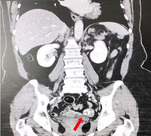
We present a case of a 58-year-old woman, G2P2, with a body mass index (BMI) of 29 kg/m2 that was referred to the Gynecology Department due to an incidental pelvic mass found on a computed tomography (CT) scan performed in the context of an acute cholecystitis (Figure 1). Her remaining medical, surgical, and family history were unremarkable, namely for ovarian or breast cancer. The physical examination and laboratory assays including tumor markers revealed no significant abnormalities.
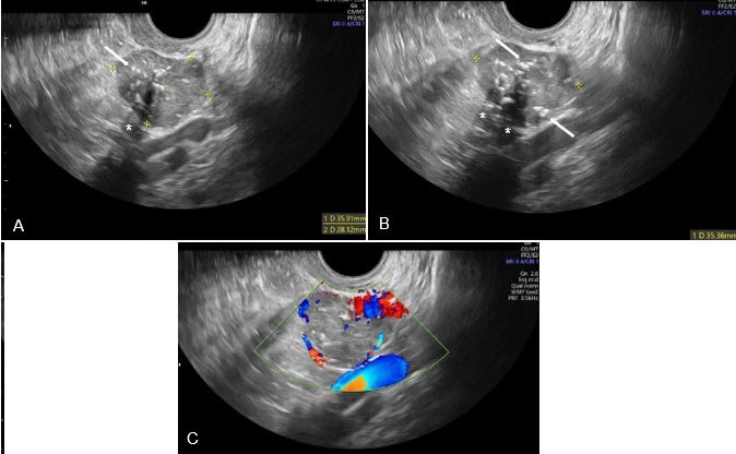
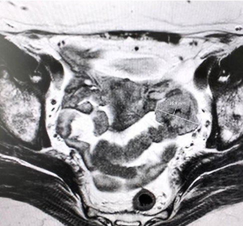
Two-dimensional ultrasonography was performed with a Voluson S10 using a 5- to 9-MHz broadband transvaginal probe. The uterus, endometrium, and ovaries appeared within normal ranges. A solid, slightly lobulated mass measuring 36 × 28 × 35 mm was visualized in the left adnexa next to the left ovary (Figure 2A and Figure 2B). Independent movement of the mass and ovary was noted when gentle pressure was applied with the transvaginal probe. The lesion exhibited a mixed echogenic pattern with scattered hyperechoic foci and multiple acoustic shadowing (Figure 2A and Figure 2B). Color Doppler was used to evaluate vascular flow, and it demonstrated moderate peripheral vascularization—color score 3/4 (Figure 2C). Based on the ultrasound images, a broad ligament fibroid was suspected but given its atypical appearance, a pelvic magnetic resonance imaging (MRI) scan was requested for better clarification. It confirmed a 36 mm solid mass in the left adnexal region, without evidence of distant involvement (Figure 3). On T1-weighted images (WI), the lesion appeared isointense relative to muscle. On T2-weighted sequences, the mass showed predominantly intermediate signal intensity, with a central area of hypointensity likely suggesting a fibrotic component. The peripheral solid component demonstrated strong diffusion restriction, appearing hyperintense on diffusion-weighted imaging (DWI) and hypointense on the apparent diffusion coefficient (ADC) map. Post-contrast T1-weighted acquisitions showed homogeneous enhancement of the solid component, while the central hypointense region did not enhance, resulting in an overall heterogeneous enhancement pattern.
After a detailed explanation and comprehensive counselling regarding the available options, the patient chose a surgical approach, and a diagnostic laparoscopy was performed. Intraoperatively, gross examination revealed a 40 × 40 mm solid mass in the left mesosalpinx, between the ovary and the fallopian tube. The tumor was well circumscribed but partially connected to the ampullary region of the left fallopian tube. The uterus, both fallopian tubes and ovaries were macroscopically unremarkable and there was no ascitic fluid. Resection with endobag extraction of the tumor mass was performed. The recovery after surgery was uneventful and the patient was discharged on the same day.
Histopathological analysis of the nodular lesion of 50 × 35 × 20 mm showed mainly tubular architectural pattern, with focal solid, retiform, and sieve-like patterns. Immunohistochemistry showed diffuse expression of pancytokeratin (AE1/AE3), calretinin and Wilms tumor 1 (WT1); multifocal expression of alfa-inhibin; focal expression of CD10; and was negative for epithelial membrane antigen (EMA), cytokeratin 7 (CK7), CD99, chromogranin, synaptophysin, GATA binding protein 3 (GATA3), and PAX8.
Histological and immunohistochemical findings supported the diagnosis of a Wolffian tumor of the broad ligament and the patient was referred to the Gynecological Oncology consultation. Considering the available information, a total abdominal hysterectomy with bilateral salpingo-oophorectomy was performed. Intraoperatively, no further pelvic or abdominal lesions were detected. The post-operative course was unremarkable, and the patient was discharged on the fourth day after surgery. In the post-operative appointment, the patient presented well and with full recovery.
DISCUSSION
The term “female adnexal tumors of probable Wolffian origin (FATWOs)” was first described by Kariminejad and Scully in 1973, who postulated its origin from adnexal mesonephric remnants [4]. The World Health Organization later classified it as Wolffian tumor, but the term FATWO remains widely used in the literature [5]. Evidence supporting its Wolffian origin includes its location within Wolffian remnants, as the broad ligament, different ultrastructural features and morphologic deviation from any neoplasms of the Müllerian or surface epithelial origin, and immunohistochemical similarities to Wolffian duct tissue [1].
While abdominal pain or an abdominal mass is a common presentation, many cases—including the one we report—are incidentally identified. The reported age range at diagnosis is 15–83 years, with an average of approximately 50 years [6],[7],[8] . Ramirez et al. reported normal preoperative serum CA-125 levels in all their patients, consistent with our findings, suggesting that CA-125 may not be a useful diagnostic marker for FATWOs [7].
Although definitive diagnosis ultimately relies on histopathology, preoperative imaging can provide valuable insight for clinical decision-making. However, there are a limited number of studies describing the ultrasound characteristics of FATWOs [2],[3] .
Female adnexal tumors of probable Wolffian origin commonly occur along the trajectory of the broad ligament, extending from the ovarian hilum to the outer third of the vagina [6] . According t oLuo and Wang, the anatomical location of the tumor in the broad ligament may serve as a useful diagnostic clue for FATWO, a feature also observed in our case. Both their report and ours describe a well-circumscribed, predominantly solid adnexal mass with moderate vascularization, though important differences exist. The authors described uniformly hypoechoic lesions, whereas our case demonstrated a mixed echogenicity with scattered hyperechoic foci and multiple acoustic shadows. Moreover, Doppler assessment in our case revealed moderate peripheral vascular flow, contrasting with the central vascularity reported by Luo et al. [2]. Our findings also partially align with those of Kuper et al., who described a similar mixed echogenic pattern and hyperechoic foci; however, unlike their case, our lesion exhibited acoustic shadowing and moderate, instead of low vascularization [3]. Although Kuper et al. mention in their discussion that acoustic shadowing is described in cases of FATWO, neither of these two studies identified this feature in their patients [2],[3]. Therefore, our work emphasizes the possibility that these tumors may exhibit acoustic shadowing, highlighting the difficulty of the differential diagnosis with leiomyoma.
Even though we believe that transvaginal ultrasound represents a crucial tool for detecting pelvic masses, distinguishing FATWO from subserosal leiomyomas or ovarian tumors can be challenging. In fact, Hong et al. have noted that the morphologic heterogeneity of FATWO poses significant diagnostic challenges, particularly in differentiating it from ovarian epithelial malignancies, sex cord-stromal tumors, and metastatic neoplasms of nongynecologic origin [9] . Rare intra-abdominal entities such as gossypiboma should also be considered in differential diagnosis, as highlighted in prior reports of incidentally detected abdominal foreign bodies on imaging [10]. Our MRI findings were consistent with previous descriptions of predominantly solid or cystic-solid adnexal tumors. On T1WI, the lesion appeared isointense to skeletal muscle, while on T2WI it demonstrated intermediate signal intensity. Additionally, increased signal on DWI and restricted ADC values within the solid components have also been described and consistent with our findings. Although some reports note a prominent low-signal-intensity rim at the tumor margin, this feature was not observed in our case [11],[12]. According to Ito et al., when a pelvic mass appears distinct from the normal ovaries, differential diagnoses include tubal carcinoma, broad ligament leiomyomas, exophytic ovarian tumors, and mesenteric gastrointestinal stromal tumors (GIST). They discuss that tubal carcinoma is typically accompanied by a dilated fallopian tube, peritoneal implants, and elevated CA-125—none of which were present in our case. Cellular leiomyomas can be difficult to distinguish, as they often show intermediate T2-weighted signal and hyperintensity on DWI, features observed here [11],[12]. Ovarian fibromas can present as exophytic masses while preserving the normal ovarian shape. Although they generally display low T2-weighted signal, degenerated fibromas may exhibit increased signal or cystic changes, complicating the differential diagnosis. Pelvic mesenteric GISTs relation with the intestine or mesenteric vessels aids diagnosis, a finding absent in this patient [11]. Hence, imaging findings were not conclusive for a specific diagnosis and cellular leiomyoma of the broad ligament remained the leading hypothesis. This underscores the importance of histological characterization.
According to Kuper et al., FATWO should be included in the differential diagnosis when sonography reveals a complex solid and cystic mass distinct from the ovary, located in the paravaginal, paraovarian, or retroperitoneal regions [3] . However, the presentation of FATWO as an adnexal mass with nonspecific clinical features makes preoperative suspicion challenging [13]. This case reinforces the need to consider FATWO as a potential diagnosis when evaluating adnexal and peri-adnexal masses. Its relevance lies in the rarity of this tumor and the limited imaging descriptions available in the literature, making each reported case a valuable contribution to improving recognition and diagnostic accuracy.
Histopathological confirmation remains crucial for establishing clinico-pathological correlations in rare conditions, reinforcing the need for careful tissue evaluation in suspected FATWO cases [14].
While a specific diagnostic marker for FATWO is lacking, immunohistochemistry provides valuable clues for distinguishing it from ovarian neoplasms [13],[15],[16]. The immunohistochemical profile observed in our study aligns closely with prior reports. Hong et al. highlighted that CD10 positivity and PAX8 negativity represent the most reliable markers for establishing the diagnosis of FATWO, which is consistent with our findings [9]. Reported CD10 expression in FATWO ranges from diffuse to focal, with a positivity rate of 50–100% [8],[9],[15],[17]. In addition to PAX8, EMA is also frequently reported as negative in FATWO [1],[8],[15],[17]. CD10 expression is particularly useful in distinguishing Wolffian tumors from sex cord-stromal neoplasms, whereas the absence of EMA expression aids in differentiating them from epithelial tumors [16]. Although CK7 is commonly reported as positive in FATWO, our case demonstrated negativity for this marker [1],[8],[15],[17]. Similarly to our case, GATA3 has been described as negative in the case series of Goyal et al., while Bennett et al. noted focal positivity for EMA, GATA3, and PAX8 in 7–20% of cases [15],[17]. Although FATWO lack a fully reproducible immunophenotypic profile, their overall immunostaining pattern remains a valuable aid in establishing the correct diagnosis, and reporting additional cases with detailed immunohistochemical profiles is essential to further refine diagnostic criteria.
Due to its rarity, there is no established consensus on the optimal management of FATWO [18]. These tumors are generally considered to have low malignant potential over time. However, recurrence and metastasis have been reported, particularly in patients who were initially treated with tumor resection alone [18],[19].
Complete surgical resection with hysterectomy, bilateral salpingo-oophorectomy and debulking surgery seems the most recommended treatment approach [16],[18]. Hence, preoperative clinical suspicion is of utmost importance to allow adequate management. While some reports suggest remission or partial response to adjuvant therapy, the efficacy of radiotherapy, chemotherapy, hormone therapy, and molecular-targeted therapy for malignant FATWO remains unclear [16]. Chemotherapy with docetaxel plus carboplatin has been proposed for recurrent and metastatic FATWOs [19].
Lesin et al. reported that the median time to recurrence was 48 months (range, 13–96 months) after treatment, based on literature review. Since in their case recurrence occurred six years after the initial treatment, they emphasize the importance of extended long-term follow-up [18]. To date, the optimal duration of surveillance has not been clearly defined in the literature.
CONCLUSION
In conclusion, the rarity of FATWO, along with nonspecific clinical manifestations, ambiguous radiologic features and variable histologic and immunohistochemical patterns makes diagnosis particularly challenging. The ultrasound characteristics of FATWOs are poorly documented in the literature with considerable variability across available descriptions. Additional case reports and case series will be essential to refine diagnostic criteria— integrating both imaging and immunohistochemical profiling features—and to facilitate the development of a more standardized therapeutic strategy, as well as structured follow-up protocols.
REFERENCES
1.
Shalaby A, Shenoy V. Female adnexal tumor of probable Wolffian origin: A review. Arch Pathol Lab Med 2020;144(1):24–28. [CrossRef]
[Pubmed]

2.
Luo J, Wang Z. The Significance of ultrasound findings in female adnexal tumor of probable wolffian origin (FATWO): Case review series. Open Journal of Obstetrics and Gynecology 2020;10:990–9. [CrossRef]
3.
Kuper SG, Wright DR, Callahan MJ, Hiett AK. Ultrasound presentation of a female adnexal tumor of probable Wolffian origin (FATWO). Journal of Diagnostic Medical Sonography 2013;29(6):282–4. [CrossRef]
4.
Kariminejad MH, Scully RE. Female adnexal tumor of probable Wolffian origin. A distinctive pathologic entity. Cancer 1973;31(3):671–7. [CrossRef]
[Pubmed]
5.
Lawrence WD, Seidman JD, Alvarado-Cabrero I, Rutgers JL, Young RH. Wolffian tumour. In: Kurman RJ, Carcangiu ML, Herrington CS, Young RH, editors. World Health Organization Classification of Tumours of Female Reproductive Organs. 4th ed. Lyon: IARC Press; 2014. p. 117–118.
6.
Wagatsuma S, Yaegashi N, Namiki T, Sato S, Yajima A. A case of female adnexal tumor of probable Wolffian origin: Histologically, tumor cells showed three different patterns. Tohoku J Exp Med 1997;181(3):371–7. [CrossRef]
[Pubmed]
7.
Ramirez PT, Wolf JK, Malpica A, Deavers MT, Liu J, Broaddus R. Wolffian duct tumors: Case reports and review of the literature. Gynecol Oncol 2002;86(2):225–30. [CrossRef]
[Pubmed]
8.
Sinha R, Bustamante B, Tahmasebi F, Goldberg GL. Malignant female adnexal tumor of probable Wolffian origin (FATWO): A case report and review for the literature. Gynecol Oncol Rep 2021;36:100726. [CrossRef]
[Pubmed]
9.
Hong S, Cui J, Li L, Buscema J, Liggins C, Zheng W. Malignant female adnexal tumor of probable Wolffian origin: Case report and literature review. Int J Gynecol Pathol 2018;37(4):331–7. [CrossRef]
[Pubmed]
10.
Kanani J, Sheikh MI. Undiagnosed, uncomplicated foreign body in abdominal cavity – A case of medical negligence. Surgery Case Reports 2024;2:100024. [CrossRef]
11.
Ito H, Koyama T, Kanie Y, Morioka K, Nakaya M, Mitsumori A, et al. MRI, CT and FDG-PET/CT findings of Wolffian tumor: Four-case series. Jpn J Radiol 2021;39(10):1009–16. [CrossRef]
[Pubmed]
12.
Cui C, Cui D, Pan J, Zhou S, Zheng X. Magnetic resonance imaging findings of a case with Wolffian tumor and related literature review. Asian Biomed (Res Rev News) 2024;18(2):81–86. [CrossRef]
[Pubmed]
13.
Amir A, Chua KJ, Varughese J. Malignant recurrence of female adnexal tumor of probable Wolffian origin (FATWO). Gynecol Oncol Rep 2020;34:100666. [CrossRef]
[Pubmed]
14.
Kanani J, Sheikh MI, Jain S, Mesuriya S. Histopathological changes in the human tissues in various types of poisoning: A cross-sectional autopsy study. Toxicol Rep 2024;13:101771. [CrossRef]
[Pubmed]
15.
Bennett JA, Ritterhouse LL, Furtado LV, Lastra RR, Pesci A, Newell JM, et al. Female adnexal tumors of probable Wolffian origin: Morphological, immunohistochemical, and molecular analysis of 15 cases. Mod Pathol 2020;33(4):734–47. [CrossRef]
[Pubmed]
16.
Rosen C, Reardon E, Shyu S, Terhune J, Saats P, Ioffe O, et al. Wolffian tumor (female adnexal tumor of Wolffian origin) presenting as a pelvic side wall mass: Report of a case. SAGE Open Med Case Rep 2019;7:2050313X19839534. [CrossRef]
[Pubmed]
17.
Goyal A, Masand RP, Roma AA. Value of PAX-8 and SF-1 immunohistochemistry in the distinction between female adnexal tumor of probable Wolffian origin and its mimics. Int J Gynecol Pathol 2016;35(2):167–75. [CrossRef]
[Pubmed]
18.
Lesin J, Forko-Ilić J, Plavec A, Planinić P. Management of Wolffian duct tumor recurrence without chemotherapy. Arch Gynecol Obstet 2009;280(5):855–7. [CrossRef]
[Pubmed]
19.
Chen Q, Shen Y, Xie C. Recurrent and metastatic female adnexal tumor of probable Wolffian origin: A case report and review of the literature. Medicine (Baltimore) 2021;100(13):e25377. [CrossRef]
[Pubmed]
SUPPORTING INFORMATION
Acknowledgments
The authors would like to thank the Gynecologic Oncology Unit, as well as the pathology and radiology departments, for their valuable contributions to the diagnosis and evaluation of this case. We also express our gratitude to the patient for consenting to share her medical information for the purpose of advancing clinical knowledge.
Generative AI technology (ChatGPT, version GPT-4, OpenAI—https://openai.com/chatgpt) was used solely for language editing and grammar correction in this manuscript. No scientific content, data analysis, or interpretation of results was generated or influenced by AI. All text was carefully reviewed and approved by the authors. The AI tool is not listed as an author, in accordance with the journal’s guidelines.
Author ContributionsRita Vasconcelos - Conception of the work, Design of the work, Analysis of data, Revising the work critically for important intellectual content, Final approval of the version to be published, Agree to be accountable for all aspects of the work in ensuring that questions related to the accuracy or integrity of any part of the work are appropriately investigated and resolved.
Diana Pereira Azevedo - Conception of the work, Design of the work, Analysis of data, Revising the work critically for important intellectual content, Final approval of the version to be published, Agree to be accountable for all aspects of the work in ensuring that questions related to the accuracy or integrity of any part of the work are appropriately investigated and resolved.
Ana Catarina Borges - Acquisition of data, Revising the work critically for important intellectual content, Final approval of the version to be published, Agree to be accountable for all aspects of the work in ensuring that questions related to the accuracy or integrity of any part of the work are appropriately investigated and resolved.
Pedro Coutinho-Borges - Acquisition of data, Revising the work critically for important intellectual content, Final approval of the version to be published, Agree to be accountable for all aspects of the work in ensuring that questions related to the accuracy or integrity of any part of the work are appropriately investigated and resolved.
Carla Monteiro - Acquisition of data, Revising the work critically for important intellectual content, Final approval of the version to be published, Agree to be accountable for all aspects of the work in ensuring that questions related to the accuracy or integrity of any part of the work are appropriately investigated and resolved.
Natacha Sousa - Conception of the work, Design of the work, Acquisition of data, Revising the work critically for important intellectual content, Final approval of the version to be published, Agree to be accountable for all aspects of the work in ensuring that questions related to the accuracy or integrity of any part of the work are appropriately investigated and resolved.
Paula Serrano - Conception of the work, Design of the work, Acquisition of data, Revising the work critically for important intellectual content, Final approval of the version to be published, Agree to be accountable for all aspects of the work in ensuring that questions related to the accuracy or integrity of any part of the work are appropriately investigated and resolved.
Guaranter of SubmissionThe corresponding author is the guarantor of submission.
Source of SupportNone
Consent StatementWritten informed consent was obtained from the patient for publication of this article.
Data AvailabilityAll relevant data are within the paper and its Supporting Information files.
Conflict of InterestAuthors declare no conflict of interest.
Copyright© 2025 Rita Vasconcelos et al. This article is distributed under the terms of Creative Commons Attribution License which permits unrestricted use, distribution and reproduction in any medium provided the original author(s) and original publisher are properly credited. Please see the copyright policy on the journal website for more information.