|
Case Report
Vulvar melanoma: A rare case report from the department of obstetrics and gynecology
1 Resident in Obstetrics and Gynecology, CHU Mohammed VI, Agadir, Morocco
Address correspondence to:
Meriem Nouri
Resident in Obstetrics and Gynecology, CHU Mohammed VI, Agadir,
Morocco
Message to Corresponding Author
Article ID: 100044G06MN2026
Access full text article on other devices

Access PDF of article on other devices

How to cite this article
Nouri M, Baloul O, El Borgi H, Kouchih A, Ammouri S, El Farouqi A. Vulvar melanoma: A rare case report from the department of obstetrics and gynecology. Edorium J Gynecol Obstet 2026;11(1):1–8.ABSTRACT
Introduction: Approximately 3% to 5% of all cancer cases in women are caused by vulvar cancer, a rare tumor compared to other gynecological tumors. It is a particularly aggressive tumor originating from melanocytes and accounts for only 2.9% of all vulvar malignancies.
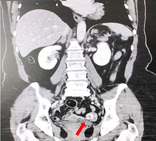
Case Report: A 56-year-old woman, single, menopausal for five years, presented with chronic left-sided vulvar pruritus evolving for approximately five years. Gynecological examination revealed a large erythematous lesion involving both labia majora, with a central erosive area and peripheral induration, associated with an irregular pigmented plaque centered on the left labium majus, extending posteriorly to the perineum and to the inner surface of the buttocks. Multiple excoriations due to scratching were noted, as well as several nevi, with no palpable inguinal lymphadenopathy. Bilateral inguinal ultrasound demonstrated lymphadenopathies of variable sizes, including nodes showing loss of fatty hilum, the largest measuring 10 × 9.4 mm on the left and 7 × 6 mm on the right, without abnormal fluid collection. Pelvic magnetic resonance imaging (MRI) revealed a vulvar tissue process located in the posterior aspect of the left labium majus, without signs of deep extension. Thoraco-abdomino-pelvic computed tomography (CT) showed no evidence of metastatic disease. The patient underwent partial vulvectomy combined with bilateral inguinal lymph node dissection followed by reconstruction using a flap procedure. Surgical margins were clear on frozen section. Final histopathological examination confirmed an invasive superficial spreading vulvar melanoma, associated with an extensive in situ component. Breslow thickness was 1 mm, with invasion limited to the superficial dermis (Chung level I). Mitotic activity was 6 mitoses per 10 high-power fields, with no ulceration, no lymphovascular invasion, and no perineural invasion. The invasive component was located more than 10 mm from all surgical margins; however, the in situ component involved the external and internal margins and was 6 mm from the superior margin. Bilateral inguinal lymph node dissection showed no metastatic involvement.
Conclusion: Vulvar melanoma is a rare and particularly aggressive tumor with a poor prognosis. Prognostic factors include tumor size, depth of invasion, tumor differentiation, surgical margins, patient age, involvement of adjacent structures, and especially lymph node status.
Keywords: Ainguinal lymphadenopathy, Case report, Vulvar melanoma
SUPPORTING INFORMATION
Author Contributions
Meriem Nouri - Substantial contributions to conception and design, Acquisition of data, Analysis of data, Interpretation of data, Drafting the article, Revising it critically for important intellectual content, Final approval of the version to be published
Oumaima Baloul - Revising it critically for important intellectual content, Final approval of the version to be published
Hajar El Borgi - Revising it critically for important intellectual content, Final approval of the version to be published
Abdillah Kouchih - Revising it critically for important intellectual content, Final approval of the version to be published
Safae Ammouri - Revising it critically for important intellectual content, Final approval of the version to be published
Abdellah El Farouqi - Revising it critically for important intellectual content, Final approval of the version to be published
Guaranter of SubmissionThe corresponding author is the guarantor of submission.
Source of SupportNone
Consent StatementWritten informed consent was obtained from the patient for publication of this article.
Data AvailabilityAll relevant data are within the paper and its Supporting Information files.
Conflict of InterestAuthors declare no conflict of interest.
Copyright© 2026 Meriem Nouri et al. This article is distributed under the terms of Creative Commons Attribution License which permits unrestricted use, distribution and reproduction in any medium provided the original author(s) and original publisher are properly credited. Please see the copyright policy on the journal website for more information.