|
Case Report
Vulvar arteriovenous malformation mimicking a Bartholin gland cyst: A case of life-threatening hemorrhage
1 MD, Resident Physician PGY3, Department of Obstetrics and Gynecology, HCA Medical City Arlington, Arlington, TX, USA
2 MD, Resident Physician PGY1, Department of Obstetrics and Gynecology, HCA Medical City Arlington, Arlington, TX, USA
3 BS, Medical Student OMS3, Department of Obstetrics and Gynecology, Texas College of Osteopathic Medicine, Fort Worth, TX, USA
4 DO, Program Director, Department of Obstetrics and Gynecology, HCA Medical City Arlington, Arlington, TX, USA
Address correspondence to:
Sandra Zhi
3201 Matlock Rd, Suite 220, Arlington, TX 78015,
USA
Message to Corresponding Author
Article ID: 100049G06RJ2026
Access full text article on other devices

Access PDF of article on other devices


How to cite this article
Johnson R, Zhi S, Khuu M, Boccaccio R. Vulvar arteriovenous malformation mimicking a Bartholin gland cyst: A case of life-threatening hemorrhage. Edorium J Gynecol Obstet 2026;11(2):30–34.ABSTRACT
Introduction: Labial masses are common and typically benign. Common causes include Bartholin cysts, sebaceous cysts, and labial warts. We report a case of a 33 year old who presented for what appeared to be a benign Bartholin gland cyst but resulted in massive hemorrhage, exam under anesthesia, massive transfusion, and embolization.
Case Report: A 33-year-old patient presents for a painless, non-enlarging labial mass. History and physical exam were consistent with a Bartholin gland cyst. Routine incision and drainage of this mass led to massive hemorrhage. The patient was emergently transferred to the operating room for an exam under anesthesia. Bleeding was controlled intraoperatively and the mass was found to be an arteriovenous malformation (AVM). The patient was stabilized and transferred to the intensive care unit (ICU). Subsequently she underwent pudendal artery embolization with interventional radiology.
Conclusion: Arteriovenous malformations are a grouping of poorly formed arteries and veins that connect to each other without a capillary bed. The prevalence of AVMs is rare and pelvic AVMs account for 2% of those instances. The majority of pelvic AVMs occur in the context of congenital conditions such as hereditary hemorrhagic telangiectasia, Cowden syndrome from PTEN (phosphatase and tensin homolog) mutations and Parkes Weber syndrome. While this AVM was visualized with doppler ultrasound, the gold standard of imaging is catheter angiography. Most vulvar AVM cases reported are in premenarchal girls with one report in a postmenopausal patient marked by pruritus over the lesion. Overall, proper history taking, physical exam, and noting of concerning features are necessary to determine the need for imaging.
Keywords: Arteriovenous malformation, Bartholin gland cyst, Clinic procedures, Incision and drainage, Massive transfusion protocol
INTRODUCTION
Labial masses are commonly identified and addressed in the clinic setting. An otherwise healthy patient complaining of a labial mass in the outpatient setting without signs of infection incur a short differential: Bartholin cysts, epidermal inclusion cysts, labial warts, sebaceous cysts, etc. The differential largely includes benign skin lesions or cysts that can be incised and drained in the clinic. However, this case describes an incident where an incision and drainage of a suspected Bartholin cyst led to a massive hemorrhage and the need for rapid escalation to a higher level of care in order to preserve life.
When a patient presents with a labial mass, there begins a simple differential. A Bartholin cyst, a benign cyst formed by the Bartholin gland at the 5 o’clock position of the labia, is a commonly suspected cause of labial masses [1]. Epidermal inclusion cysts are the most common cutaneous cyst and can occur anywhere on the body [2]. Labial warts can also be a benign finding caused by human papillomavirus types 6 and 11 acquired through sexual contact [3]. Sebaceous cysts can also present in the labia, which are benign keratin-filled cysts that grow slowly over time [4]. Leiomyoma can present in the labia as well. Some rarer conditions can include hematomas, hernias, angiomyxoma, and many other possibilities. Our case presents a patient with a labial mass that truly masquerades as a well-circumscribed Bartholin cyst and its true identity is not discovered until after the patient had a life-threatening complication.
CASE REPORT
A 33-year-old G2P2002 patient presented to the clinic with a complaint of a “vaginal bump,” which she had first noticed three weeks before presentation. In that time, it had not significantly grown. It was not painful but mildly uncomfortable when it was palpated. She denies changes to her urination, sexual function, and menstruation. Her medical history was also notable for difficult to control hypertension, seizures of unknown etiology, migraines, and premature ventricular contractions. She had been on and off several medications in an attempt to control her blood pressures including various antihypertensives and midodrine, a medication to increase blood pressure. She was not taking any antihypertensive medications at the time of her presentation. She did have a relative with sudden cardiac death. She denied any history of sexually transmitted diseases.
On presentation her blood pressure was 160/97 with a repeat of 140/100. Her physical exam was normal except for a 1.5 cm left cystic mass at the 5 o’clock position of her labia. There were no overlying skin changes. It was nontender and nonpulsatile. The patient was counseled on incision and drainage of suspected Bartholin’s gland cyst. The overlying skin was cleansed with povidone and numbed with lidocaine with epinephrine. After a few minutes the patient described a tingling in her jaw and ears but was otherwise unchanged. The mass was palpated, and an 11 blade was used to incise the mass. Brisk bloody fluid was produced from the incision site. The flow was constant and did not appear pulsatile. Initially it was suspected to be bloody fluid from a cyst. However, the fluid continued to be brisk and bloody after 3–5 seconds. Additional office personnel were called for. An attempt was made to pack the area but was unsuccessful and bleeding continued briskly. The adjacent hospital operating room was called and an emergent case for an exam under anesthesia was ordered. Blood products were ordered as well. The patient was transferred to a wheelchair and emergently wheeled to the operating room by the physicians taking care of her.
During transit the patient is estimated to have lost approximately 2 liters of blood in approximately 15 minutes. Upon arrival at the operating room (OR) she developed an altered level of consciousness. She no longer responded to commands and had to be lifted by OR personnel onto a gurney. One of the physicians then held pressure on the mass while the patient was wheeled to the OR. She was slid to the OR table and was splash prepped with Betadine and general anesthesia was induced without difficulty. Two 0 Vicryl pops were placed in figure-of-eight fashion to achieve hemostasis. The suspected Bartholin’s cyst was still felt to be intact so an additional incision with an 11 blade was created to drain the cyst; however, upon the second incision similar bleeding resumed. Hemostasis was achieved at this point also with two 0 Vicryl figure-of-eight sutures. An ultrasound probe was placed on the area that revealed a cystic dilated structure with arterial and venous blood flow consistent with an arteriovenous malformation (AVM). The defect was not growing in size and hemostasis had been achieved vaginally. The patient was packed with a Kerlix packing and Foley was placed. She was transferred to the intensive care unit (ICU) with plans to consult interventional radiology. She received four units of packed red blood cells and four units of fresh frozen plasma as well as 500 cc of lactated ringers. Perioperative hemoglobin and hematocrit was not collected due to the emergent nature of the event; however, levels one month prior had been 13.8/44.4. Immediately postoperative levels were 10.1/31.
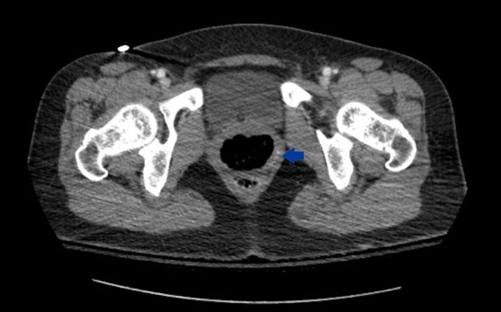
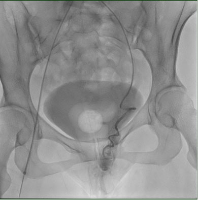
Once in the ICU the patient was able to be extubated but required a nicardipine drip to maintain her blood pressure. This was discontinued on postoperative day one and she subsequently needed carvedilol 6.25 twice a day and amlodipine 10 mg daily. Computed tomography (CT) angiography scan showed an asymmetric single tortuous arterial vessel in the left vaginal wall. Hereditary hemorrhagic telangiectasia was considered as a possible diagnosis but there was no family history that would support this diagnosis. While a solitary AVM does not increase the likelihood of cerebral AVMs, her history of seizure of unknown etiology did raise concern for one. A CT head and neck showed no abnormal vasculature. On postoperative day one interventional radiology successfully embolized the AVM originating from the left pudendal artery with Gelfoam slurry. The patient was downgraded from the ICU and discharged on postoperative day two (Figure 1 and Figure 2).
DISCUSSION
Arteriovenous malformations are a grouping of poorly formed arteries and veins that connect to each other without a capillary bed. These arteries have dysfunctional muscular layers and without the pressure buffer from the capillary beds, the veins are often blown out and dilated [5]. Arteriovenous malformations are categorized as fast-flow vascular malformations and carry significant bleeding risks when disturbed. Causes of arteriovenous malformations remain unclear but are thought to be due to angiogenic stimulation from pre-existing vessels and certain genetic mutations [5].
Pelvic arteriovenous malformations are rare and account for less than 2% of all reported AVMs [6]. Unlike other malformations, pelvic AVMs show a preference for sex, with 68% of pelvic arteriovenous malformations occurring in women [6]. They also tend to be more complex when found in women; on average, female malformations have more feeder arteries than males. Symptoms caused by AVMs are broad and mostly dependent on their location, though 20% of AVMs remain asymptomatic [7]. Superficial malformations may be palpable, feeling soft and spongy in texture and may increase in size with changes in position [7].
Multiple cases of vulvar AVMs have been documented to present as an inflamed, ulcerated, tender, swollen, and nodular mass, similar to how a Bartholin’s cyst may be described [8],[9],[10]. It is important to consider rare causes of vulval lesions prior to operative procedures of suspected benign pathologies. All AVMs carry a hemorrhage risk but vulvar lesions are located in an area with a high concentration of important vasculature and nerves [11]. Misdiagnosis could have been avoided if imaging had been done prior to the procedure.
The best method for AVM visualization is catheter angiography, but due to its invasiveness, it is reserved until treatment is administered. Treatment often requires medical and surgical approaches. Medical treatment includes mammalian target of rapamycin (mTOR) inhibitors, β blockers, and anti-androgens [8]. Surgical treatment ranges from embolization to complete resection. Recurrence occurs more frequently in women due to their tendency to have multiple feeder arteries. On average, female patients required 3.2 rounds of embolization compared to males who only had 1.7 average over a 23.3 month period [8].
Most vulvar AVM cases described in literature are in premenarchal girls, suggesting a congenital cause of the AVMs [12]. One such case report presents a 16-year-old female with a large, painful, and nodular mass that was stable for two years prior but now became bothersome in symptoms [11]. Our patient presented after noticing the mass, but did not have any overlying skin changes. It is possible that the mass could have mirrored those symptoms with time as AVMs will continue to grow if not treated. An interesting feature of this case described above is that this patient has had two vaginal deliveries in the past without significant hemorrhage, noting that this mass remained undiscovered during her pregnancies. Another case describes a 57-year-old postmenopausal woman with a vulvar mass with overlying pruritus [13], which is a different presentation compared to our patient and warrants an initial evaluation for vulvar cancer. Ultimately, our patient presents a terrifying scenario when a very benign presentation results in a hemorrhage in a setting where the providers are not routinely prepared to tamponade such bleeding.
CONCLUSION
Given this unexpected critical situation in the setting of a presumed benign outpatient procedure, it will be critical to correctly identify this aberrant pathology. In order to assess if a mass needs to be imaged as a part of its evaluation, a full history and physical exam must be taken. A patient with a personal history of vessel disorders should raise suspicion for possible hematological abnormalities that can present as masses. A physical examination should include noting lesion morphology, number, size, demarcation, skin changes overlying lesion, color, consistency, texture, and any secondary characteristics such as presence of excoriation or edema, and any acute inflammation. More concerning features of a labial mass include masses that have presented in faster timelines or are associated with redness, sudden swelling, skin discoloration, or fixed masses. A pulsating mass or blue-purple discoloration would warrant further evaluation with Doppler ultrasound. Ultimately, the patient above had presented with a relatively simple-appearing cyst that was nonpulsatile and nontender with no overlying skin changes. There were no suggestive features of the lesion that would indicate a need for imaging or ultrasound. Given that the rarity of pelvic arteriovenous malformations, routine ultrasound examination of the lesions would not be recommended.
This case also brought to light the challenges involved with transitioning an office patient to a hospital patient in such a critical situation. This patient was physically placed on a wheelchair with an attempt to pack the wound and immediately wheeled to the operating room. There were challenges with activating Massive Transfusion Protocol as the patient had not been properly registered upon presentation to the OR. After a debrief with staff, an alternative was moving the patient directly to the emergency room as a trauma patient. The advantages of this decision would be to start lifesaving measures such as Massive Transfusion Protocol earlier. This incident has sparked a larger conversation at our facility about how to expedite patient care from the office setting to the operating room. Though situations like these are exceedingly rare, crucial seconds and minutes that can be saved with a standard protocol in place can alter outcomes.
REFERENCES
1.
Carlson K, Wittler M. Bartholin gland cyst. In: StatPearls. Treasure Island (FL): StatPearls Publishing; 2025.
[Pubmed]

2.
Weir CB, Sathe NC, St. Hilaire NJ. Epidermal inclusion cyst. In: StatPearls. Treasure Island (FL): StatPearls Publishing; 2026.
[Pubmed]
3.
Aguilera-Barrantes I, Magro C, Nuovo GJ. Verruca vulgaris of the vulva in children and adults: A nonvenereal type of vulvar wart. Am J Surg Pathol 2007;31(4):529–35. [CrossRef]
[Pubmed]
4.
Zito PM, Scharf R. Epidermoid cyst (archived). In: StatPearls. Treasure Island (FL): StatPearls Publishing; 2023.
[Pubmed]
5.
Bokhari MR, Bokhari SRA. Arteriovenous malformation of the brain (archived). In: StatPearls. Treasure Island (FL): StatPearls Publishing; 2023.
[Pubmed]
6.
Izumi T, Osawa M, Tsuru I, Ono A, Kume H, Shiga Y, et al. A giant pelvic arteriovenous malformation. IJU Case Rep 2024;7(5):399–403. [CrossRef]
[Pubmed]
7.
Jacobowitz GR, Rosen RJ, Rockman CB, Nalbandian M, Hofstee DJ, Fioole B, et al. Transcatheter embolization of complex pelvic vascular malformations: Results and long-term follow-up. J Vasc Surg 2001;33(1):51–5. [CrossRef]
[Pubmed]
8.
Vienet-Legué L, Hersant B, Bisdorf A, Aymard A, Owen C, Théodore C, et al. Vulvar arteriovenous malformation in a 16-year-old girl. J Pediatr Adolesc Gynecol 2018;31(1):58–61. [CrossRef]
[Pubmed]
9.
Brotherton J, Yazdany T. Resection of a vulvar arteriovenous malformation in a premenarchal patient. Obstet Gynecol 2010;115(2 Pt 2):426–9. [CrossRef]
[Pubmed]
10.
Revicky V, Maina W, Cockburn J, Stanley K. Vulval low-flow arteriovenous malformation. Arch Gynecol Obstet 2009;280(2):271–3. [CrossRef]
[Pubmed]
11.
Ku L, Ma X. Congenital giant pelvic arteriovenous malformation. Am J Med Sci 2025;369(2):e2–3. [CrossRef]
[Pubmed]
12.
Focseneanu MA, Merritt DF. Vulvar venous malformations in premenarchal girls: A report of four cases. J Pediatr Adolesc Gynecol 2012;25(2):e49–55.
13.
Pereira N, Dormosh M, Mirmanesh M, Guilfoil DS. Arteriovenous malformation of the vulva: A case report. J Low Genit Tract Dis 2014;18(1):E12–5. [CrossRef]
[Pubmed]
SUPPORTING INFORMATION
Acknowledgments
Thank you Dr. Michelle McCarroll, PhD and Hannah Affleck who provided critical assistance in guiding us throughout the submission process and investigative writing.
Author ContributionsRebecca Johnson - Conception of the work, Design of the work, Drafting the work, Revising the work critically for important intellectual content, Final approval of the version to be published, Agree to be accountable for all aspects of the work in ensuring that questions related to the accuracy or integrity of any part of the work are appropriately investigated and resolved.
Sandra Zhi - Conception of the work, Design of the work, Drafting the work, Revising the work critically for important intellectual content, Final approval of the version to be published, Agree to be accountable for all aspects of the work in ensuring that questions related to the accuracy or integrity of any part of the work are appropriately investigated and resolved.
Megan Khuu - Drafting the work, Revising the work critically for important intellectual content, Final approval of the version to be published, Agree to be accountable for all aspects of the work in ensuring that questions related to the accuracy or integrity of any part of the work are appropriately investigated and resolved.
Robert Boccaccio - Conception of the work, Design of the work, Revising the work critically for important intellectual content, Final approval of the version to be published, Agree to be accountable for all aspects of the work in ensuring that questions related to the accuracy or integrity of any part of the work are appropriately investigated and resolved.
Guaranter of SubmissionThe corresponding author is the guarantor of submission.
Source of SupportNone
Consent StatementWritten informed consent was obtained from the patient for publication of this article.
Data AvailabilityAll relevant data are within the paper and its Supporting Information files.
Conflict of InterestAuthors declare no conflict of interest.
Copyright© 2026 Rebecca Johnson et al. This article is distributed under the terms of Creative Commons Attribution License which permits unrestricted use, distribution and reproduction in any medium provided the original author(s) and original publisher are properly credited. Please see the copyright policy on the journal website for more information.