|
Case Report
Antenatal diagnosis and EXIT procedure management of an oropharyngeal fetus in fetu: A rare case report
1 Consultant, Fetal and Maternal Medicine Unit, Royal Hospital, Muscat, Oman
2 Consultant, Obstetric Ultrasound Unit, Royal Hospital, Muscat, Oman
3 Resident, Oman Medical Specialty Board, Muscat, Oman
Address correspondence to:
Bashair Al Siyabi
Resident, Oman Medical Specialty Board, Muscat,
Oman.
Message to Corresponding Author
Article ID: 100048G06BS2026
Access full text article on other devices

Access PDF of article on other devices


How to cite this article
Al Saadi B, Al Ghailani A, Al Kalbani A, Al Siyabi B. Antenatal diagnosis and EXIT procedure management of an oropharyngeal fetus in fetu: A rare case report. Edorium J Gynecol Obstet 2026;11(2):24–29.ABSTRACT
Introduction: Fetus in fetu (FIF) is an extremely rare congenital anomaly with a reported incidence of 1 in 500,000 live births. Histologically, it is characterized by a calcified vertebral axis with variably differentiated organ systems and limbs. Although the majority of FIF cases arise within the retroperitoneum, we present a rare case of an oropharyngeal FIF detected on antenatal ultrasonography.
Case Report: A 23-year-old primigravida was referred at 23 weeks of gestation following routine anomaly scanning that identified a complex oropharyngeal mass. Subsequent antenatal ultrasonography and magnetic resonance imaging (MRI) demonstrated progressive mass enlargement causing oral cavity obstruction and severe polyhydramnios. The patient underwent emergency cesarean section with ex utero intrapartum treatment procedure (EXIT). A gross examination of the new born confirmed the presence of a FIF mass with identifiable lower limbs and fetal structures.
Conclusion: Despite the rarity of a FIF mass, such cases are still encountered. The oropharyngeal location of this mass adds up to the rarity of this condition as most the reported cases are retroperitoneal. This highlights the importance of a detailed antenatal assessment aids in the management and counseling.
Keywords: Epignathus, EXIT procedure, Fetus in fetu, Oropharyngeal mass, Teratoma
INTRODUCTION
Fetus in fetus is a very rare congenital anomaly with a prevalence of 1 in 500,000 cases [1]. In this condition a fetus incorporates the well differentiated tissue of its co-twin. The vertebral column is the commonly reported component and it is a distinguishable feature between a teratoma and a fetus in fetu (FIF) mass [2]. The initial reporting of such cases dates back to 18th century by Johann Friedrich Meckel [3]. While the majority of masses a rise in the retroperitoneal space, we report a rare case of FIF arising from the oral cavity. The case was originally identified in the 23 weeks anatomy scan and was then followed antenatally and postnatally. The advancement in ultrasound and prenatal diagnostic testing aids in the planning and management of such case.
CASE REPORT
Patient Information and Antenatal History
A 23-year-old primigravida (non-consanguineous marriage and spontaneous conception) was referred to the Fetal Medicine Clinic at 23 weeks of gestation following an abnormal routine anomaly scan. Her past medical, surgical, and gynecological histories were unremarkable. She was medically free of chronic illnesses, non-diabetic, non-hypertensive, and fully vaccinated.
Initial Diagnostic Assessment
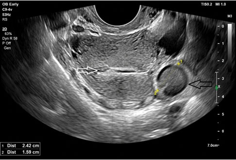
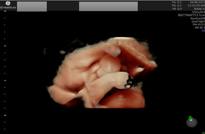
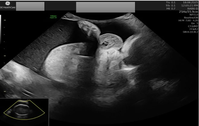
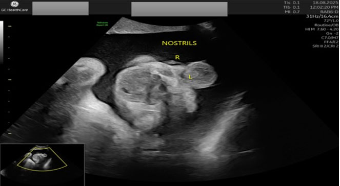
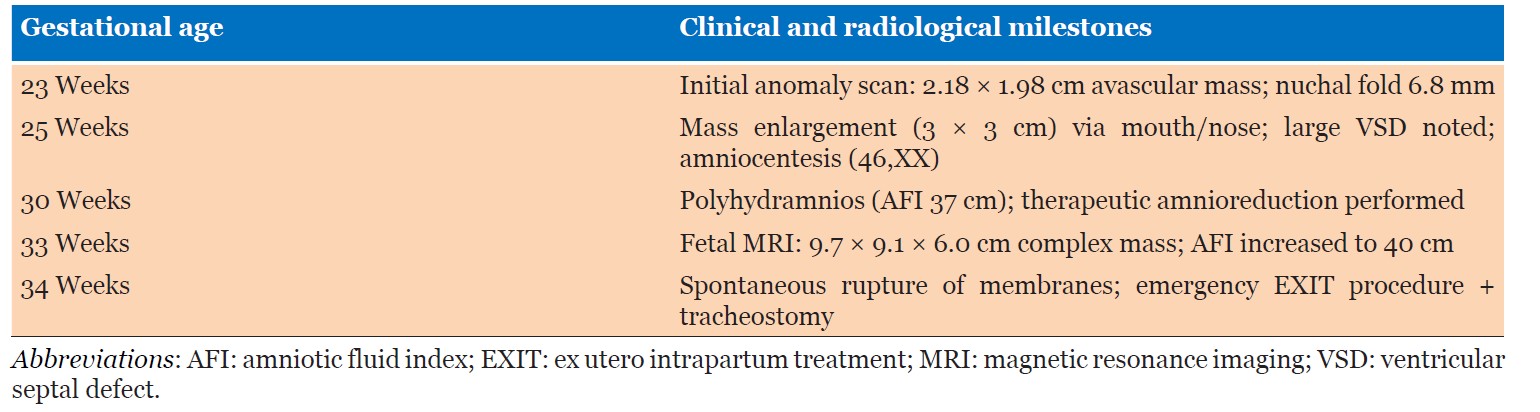
The initial 23-week anatomy scan revealed an avascular mass measuring 2.18 × 1.98 cm with an increased nuchal fold thickness of 6.8 mm. Follow-up ultrasonography at 25 weeks demonstrated a complex, avascular mass measuring approximately 3.0 × 3.0 cm originating from the pharynx and protruding through the oral and nasal cavities (Figure 1). Disruption of the hard palate was noted, and a cleft palate could not be ruled out (Figure 2 and Figure 3). Fetal echocardiography at 25 weeks revealed cardiomegaly with a large ventricular septal defect (VSD). Fetal biometric parameters were appropriate for gestational age. Differential diagnoses included pharyngeal hemangioma, teratoma, or congenital malignancy. Amniocentesis performed at 25 weeks confirmed a normal female karyotype (46,XX) with quantitative fluorescent polymerase chain reaction (QF-PCR) negative for trisomies 13, 15, 16, 18, 21, and 22. The patient was counseled regarding the guarded prognosis of the fetal condition. A multidisciplinary team discussion involving a fetal medicine consultant, a neonatologist, an anesthetist, and a pediatrician was conducted with both parents. Since local regulations do not permit termination beyond 19 weeks of gestation, and given that the prognosis was guarded with no clear antenatal diagnosis, termination was not offered to the couple. Furthermore, both parents were fully committed to the pregnancy and wished to proceed with any intervention that might save their baby’s life.
Therapeutic Interventions and Delivery
Due to progressive oral cavity obstruction preventing fetal swallowing, the patient developed severe polyhydramnios [amniotic fluid index (AFI) 37 cm] requiring therapeutic amnioreduction at 30 weeks.
Fetal magnetic resonance imaging (MRI) at 33 weeks demonstrated a large, pedunculated complex soft-tissue mass with solid and cystic components originating from the pharynx, measuring 9.7 × 9.1 × 6.0 cm, completely obstructing the upper airway. Follow-up ultrasonography showed persistent severe polyhydramnios (AFI 40 cm).
A multidisciplinary team comprising fetal medicine specialists, neonatologists, pediatric ear, nose, and throat (ENT) surgeons, pediatric cardiologists, and anesthesiologists planned an ex utero intrapartum treatment (EXIT) procedure via elective cesarean section. However, at 34 weeks of gestation, the patient presented with spontaneous rupture of membranes and active preterm labor.
An emergency cesarean section with an EXIT procedure was performed. A live female neonate weighing 2.1 kg was delivered in a cephalic presentation (APGAR scores 6 and 8 at 1 and 5 minutes, respectively). Intraoperatively, marked abdominal wall vascularity and massive polyhydramnios were noted. While maintained on uteroplacental support during the EXIT procedure, an immediate tracheostomy was secured by the ENT team (Table 1).
Postnatal Course and Outcome
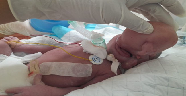
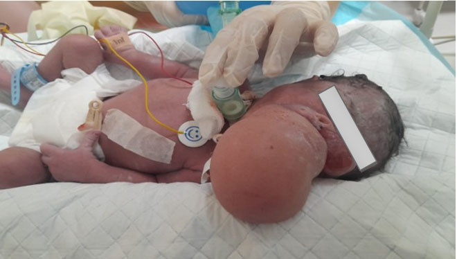
The neonate was transferred to the neonatal intensive care unit (NICU). Gross macroscopic inspection of the protruding mass confirmed well-formed fetal structures, including distinct lower limbs (Figure 4 and Figure 5). Postnatal neck soft-tissue MRI confirmed an epignathus-type FIF arising from the oropharynx causing complete mechanical airway obstruction. The mass contained differentiated neural, adipose, osseous, and soft tissues without intracranial extension.
During her 45-day NICU stay, the neonate was managed for concurrent medical issues: a large VSD, large patent ductus arteriosus (PDA), neonatal sepsis, pneumothorax, and intraventricular hemorrhage. A multidisciplinary decision (neonatology, hematology, cardiology, ENT, and pediatric surgery) was made to delay definitive surgical excision until the infant reached hemodynamic stability and developmental maturity.
DISCUSSION
Fetus is fetus (FIF) is an extremely rare condition with a reported incidence of 1 in 500,000 live births [1]. It was first described by Willis who defined it as a mass containing a calcified vertebral axis with the presence of other fetal parts or limbs seen growing as a parasitic structure in another full grown fetus. Hence the name fetiform mass [3]. There are so far two theories which are proposed to explain this condition, teratoma theory and Identical twin theory. The teratoma theory refers to a very specialized teratoma which contains highly organized tissue and mature organs. This teratoma usually appears at the same time of FIF occurrence. The identical twin theory refers to abnormal embryogenesis in the early stages of fetal development (blastocyst stage). The rapidly growing totipotent cells within the blastocyst develop into a cluster of cells that eventually develops into a fully grown fetus if it gets enough placental blood flow. Due to the lack of sufficient blood supply, some of these clusters will cease to grow and remain underdeveloped and encased within the normally growing co-twin [1]. An attempt was made to differentiate between a teratoma and a FIF. Teratomas do not usually demonstrate vertebrae or systematic organogenesis and they are commonly seen in the sacrococcygeal region. They may also have malignant potentials. On the other hand, FIF is a benign mass with a vertebral skeleton and variably differentiated organ system and limbs [4].
There are no unified clear diagnostic terms for FIF, while the presence of a macroscopic/microscopic vertebrae was necessary for the diagnosis, a lot of cases in the English literature have departed from this definition to include cases with no identifiable vertebral bodies [3]. In fact the most reported organ present within FIF mass is the vertebral column 91% followed by 82.5% limbs, 55.8% central nervous system (always anencephalic), 45% gastrointestinal tract, 40% vessels, and 26.5% genitourinary tract. To date, the heart was rarely found in FIF (about 1–2 reported cases) [2]. The absence of the cardiovascular system has led to misdiagnosis of a cardiac twin due to similar findings with FIF [5]. However, while the karyotype in FIF is normal or similar to his host twin, about 50% of a cardiac twin are associated with abnormal karyotype including both trisomy and triploidy [6]. In our reported case, the result of amniocentesis was normal female karyotype. Despite the fact that the neonate did not undergo immediate surgical excision of the mass post-delivery, macroscopic and radiological findings confirmed the diagnosis of FIF mass.
Most cases of FIF occur during infancy and the oldest reported case was of a 47-year-old man [5]. The majority of masses are seen in the retroperitoneal space which makes about 80% of the cases [7]. In a systematic review involving all reported cases of FIF till 2023, the retroperitoneum (72.2%) was the most common location, followed by intraperitoneal sites (5.8%). Other less commonly reported sites include intrathoracic (4.7%), head-and-neck region including oral cavity (4.1%), sacrococcygeal/gluteal/perineal (4.1%), intracranial (3.7%), and within the gonads (3.7%) [3]. As in our reported case, the mass originated from the oropharynx, which is an unusual site, and the cases we found in literature were only of immature teratomas.
The typical symptoms of such FIF is usually the result of a mass effect on adjacent organs. These include abdominal distension, emesis, feeding difficulty, failure to thrive, jaundice, and pressure effects in the renal or the gastrointestinal tract [4]. In fact, abdominal pain was seen in about 70% of reported cases [8]. The mass-like structure may contain a single or multiple fetuses attached by an umbilical cord with or without placenta [5]. In a case report of FIF, a two month Chinese male infant with intrapelvic mass containing three fetuses [6]. The maximum number of reported FIF in a single host till date is 9 [8].
Imaging plays a vital role in the initial evaluation of FIF masses [9]. A plain abdominal X-ray might help to identify the presence of a vertebral column and an axial skeleton as half of the reported cases may show the prior two structures [10],[11],[12]. Ultrasonographic features may show the presence of an ill-defined mass with complex solid components. In fact, ultrasound examination is considered safe and convenient for prenatal and antenatal diagnosis. However, due to the similar sonographic features between a teratoma and FIF, ultrasound can be misleading in the diagnosis [1]. Other imaging modalities like computed tomography (CT) and MRI may guide preoperative diagnosis [8]. The non-visualization of the vertebral axis in the radiography and CT does not exclude the diagnosis of fetus in fetus [5]. In our reported case, all imaging modalities have been utilized to define the nature of the mass.
Tumor markers including alpha fetoprotein (AFP), carcinoembryonic antigen test (CEA), and beta-human chorionic gonadotropin (β-hCG) were incorporated in the preoperative work-up of patients with FIF. However, a positive association between FIF and aforementioned tumor markers was lacking. Therefore, the level of the tumor markers is not used to rule in or out the diagnosis of FIF [3]. None of the tumor markers were requested in our reported case.
Complete surgical excision of FIF mass is the gold standard treatment once the diagnosis is confirmed. It is associated with a good prognosis and excellent quality of life [13]. The success of the surgical procedure is dependent on the size, location, local pressure, intracystic mass bleeding, and other uncertain diagnosis [1]. A close postoperative monitoring with imaging and tumor markers was proposed. Despite that most components of FIF mass are mature, malignant transformation after FIF mass resection was reported. Hopkins et al. reported a case of malignant transformation after surgical resection of FIF mass. Persistent elevated tumor markers such as AFP and β-hCG can be a sign of malignant transformation [10].
CONCLUSION
Oropharyngeal fetus in fetu is an exceptionally rare congenital malformation. Comprehensive antenatal imaging using high-resolution ultrasonography and fetal MRI is vital for accurate diagnosis and for facilitating advanced delivery strategies—such as the EXIT procedure—to prevent acute neonatal airway obstruction.
REFERENCES
1.
Xiaowen M, Lingxi C, Song L, Shengbao P, Xiaohong Y, Xinghai Y. Rare fetus-in-fetu: Experience from a large tertiary pediatric referral center. Front Pediatr 2021;9:678479. [CrossRef]
[Pubmed]

2.
Brand A, Alves MC, Saraiva C, Loío P, Goulão J, Malta J, et al. Fetus in fetu—diagnostic criteria and differential diagnosis—A case report and literature review. J Pediatr Surg 2004;39(4):616–8. [CrossRef]
[Pubmed]
3.
Palo S, Mangla M, Gabbeta S, Motwani R. Demystifying fetus-in-fetu: A systematic review of its clinical and pathological attributes. J Indian Assoc Pediatr Surg 2024;29(5):406–16. [CrossRef]
[Pubmed]
4.
Rai R, Singh DK, Agrahari D, Kamra P. Double fetus in fetu. Indian Pediatr 2011;48(11):904–6.
[Pubmed]
5.
Hoeffel CC, Nguyen KQ, Phan HT, Truong NH, Nguyen TS, Tran TT, et al. Fetus in fetu: A case report and literature review. Pediatrics 2000;105(6):1335–44. [CrossRef]
[Pubmed]
6.
Hing A, Corteville J, Foglia RP, Bliss DP Jr, Donis-Keller H, Dowton SB. Fetus in fetu: Molecular analysis of a fetiform mass. Am J Med Genet 1993;47(3):333–41. [CrossRef]
[Pubmed]
7.
Willis RA. The borderland of embryology and pathology. Bull N Y Acad Med 1950;26(7):440–60.
[Pubmed]
8.
Knox AJ, Webb AJ. The clinical features and treatment of fetus in fetu: Two case reports and a review of the literature. J Pediatr Surg 1975;10(4):483–9. [CrossRef]
[Pubmed]
9.
Gerber RE, Kamaya A, Miller SS, Madan A, Cronin DM, Dwyer B, et al. Fetus in fetu: 11 fetoid forms in a single fetus: Review of the literature and imaging. J Ultrasound Med 2008;27(9):1381–7. [CrossRef]
[Pubmed]
10.
Luzzatto C, Talenti E, Tregnaghi A, Fabris S, Scapinello A, Guglielmi M. Double fetus in fetu: Diagnostic imaging. Pediatr Radiol 1994;24(8):602–3. [CrossRef]
[Pubmed]
11.
Gangopadhyay AN, Srivastava A, Srivastava P, Gupta DK, Sharma SP, Kumar V. Twin fetus in fetu in a child: A case report and review of the literature. J Med Case Rep 2010;4:96. [CrossRef]
[Pubmed]
12.
Taher HMA, Abdellatif M, Wishahy AMK, Waheeb S, Saadeldin Y, Kaddah S, et al. Fetus in fetu: Lessons learned from a large multicenter cohort study. Eur J Pediatr Surg 2020;30(4):343–9. [CrossRef]
[Pubmed]
13.
Hopkins KL, Dickson PK, Ball TI, Ricketts RR, O’Shea PA, Abramowsky CR. Fetus-in-fetu with malignant recurrence. J Pediatr Surg 1997;32(10):1476–9. [CrossRef]
[Pubmed]
SUPPORTING INFORMATION
Acknowledgments
A gesture of gratitude from all the authors to the patients legal guardians for accepting to write and publish the case.
Use of Artificial Intelligence
Gemini 3.6 Flash model was used for editing and proofreading of the introduction and the discussion sections.
Badriya Al Saadi - Acquisition of data, Analysis of data, Revising the work critically for important intellectual content, Final approval of the version to be published, Agree to be accountable for all aspects of the work in ensuring that questions related to the accuracy or integrity of any part of the work are appropriately investigated and resolved.
Amira Al Ghailani - Acquisition of data, Revising the work critically for important intellectual content, Final approval of the version to be published, Agree to be accountable for all aspects of the work in ensuring that questions related to the accuracy or integrity of any part of the work are appropriately investigated and resolved.
Ahlam Al Kalbani - Acquisition of data, Revising the work critically for important intellectual content, Final approval of the version to be published, Agree to be accountable for all aspects of the work in ensuring that questions related to the accuracy or integrity of any part of the work are appropriately investigated and resolved.
Bashair Al Siyabi - Acquisition of data, Drafting the work, Revising the work critically for important intellectual content, Final approval of the version to be published, Agree to be accountable for all aspects of the work in ensuring that questions related to the accuracy or integrity of any part of the work are appropriately investigated and resolved.
Guaranter of SubmissionThe corresponding author is the guarantor of submission.
Source of SupportNone
Consent StatementAn informed consent for publication of the case with the ultrasound images was obtained from both parents who are the legal guardians of the fetus.
Data AvailabilityAll relevant data are within the paper and its Supporting Information files.
Conflict of InterestAuthors declare no conflict of interest.
Copyright© 2026 Badriya Al Saadi et al. This article is distributed under the terms of Creative Commons Attribution License which permits unrestricted use, distribution and reproduction in any medium provided the original author(s) and original publisher are properly credited. Please see the copyright policy on the journal website for more information.