 |
Research Articles
Uterine sarcomas in Dakar: Epidemiological and histopronostic aspects of a Senegalese study of 17 cases
1 Laboratory of Pathological Anatomy and Cy-tology of Thiès Regional Hospital, Avenue Malick Sypro-longée; B.P 34A Thiès RP, Senegal
2 University Hospital Aristide le Dantec, Pathology Anatomy and Cytology Department, Avenue Pasteur, BP Dakar, Senegal
Address correspondence to:
Tonleu Linda Bentefouet
Senior Lecturer, Training and Research Unit (UFR), for Health Sciences BP: 22 RP Thiès,
Senegal
Message to Corresponding Author
Article ID: 100022G06TB2019
Access full text article on other devices

Access PDF of article on other devices


How to cite this article
Bentefouet TL, Gaye AM, Thiam I, Hatim J. Uterine sarcomas in Dakar: Epidemiological and histopronostic aspects of a Senegalese study of 17 cases. Edorium J Gynecol Obstet 2019;5:100022G06TB2019.ABSTRACT
Aims: Uterine sarcomas account for about 1% of all malignant tumors of the female genital tract. They are rare tumors characterized by a great histological heterogeneity and a bad prognosis. The aim of this work was to describe the epidemiological profile and histopronostic characteristics of uterine sarcomas diagnosed in the pathological anatomy and cytology laboratories of the hospitals of Dakar.
Methods: We have conducted a retrospective and descriptive study of the uterine sarcomas over a period of 10 years. Patients were regrouped from the histological reports of the anatomy and cytology laboratories of Dakar. All patients with a histological diagnosis of uterine sarcomas were included. For each of the patients, special interest was in epidemiological and histopronostic aspects.
Results: Seventeen (17) patients were identified. All patients were of black race, 82.3 of whom were in a postmenopausal stage. The average age was 56.3 years. Leiomyosarcoma was the most common histological type, followed by endometrial stromal sarcoma, carcinosarcoma, and adenosarcoma, respectively. An immunohistochemical study was performed for seven patients.
Conclusion: Immunohistochemistry, although rarely practiced in our laboratory, is necessary for a better characterization of uterine sarcomas.
Keywords: Immunohistochemistry, Pathology anatomy, Senegal, Uterine sarcoma
INTRODUCTION
Uterine sarcomas are rare tumors. They represent 3 to 7% of cancers of the uterus [1], and are characterized by great histopathological heterogeneity. The histopathological diversity of these lesions has made it difficult to establish a consensus that allows for a good coding of their prognostic factors and their therapeutic treatment [1]. Uterine sarcomas have been poorly studied despite being observed in practice in the various pathological anatomy and cytology laboratories (ACPs). Through this work, we propose to describe the epidemiological profile and histopronostic characteristics of uterine sarcomas diagnosed in ACP laboratories in Dakar.
MATERIALS AND METHODS
This is a retrospective and descriptive study of uterine sarcomas collected in ACP laboratories of Dakar hospitals over a period of 10 years (January 2004 to December 2013). This study was based on the exploitation of the anatomopathological reports of the ACP laboratories of Aristide le Dantec Hospital (HALD), Cheikh Anta Diop University, Grand-Yoff General Hospital (Hoggy), and of Hopital Principal de Dakar (HPD), a Military Training hospital. All patients with uterine sarcoma performed on operated specimens were included, and a survey specifying the epidemiological aspects (age, race, and menopausal status) and histopronostic factors (histological type, tumor size, myometrial invasion, and presence or non-presence of vascular emboli) of each patient was completed. We carried out a rereading of observed cases after making new slides and new hematoxylin eosin coloring from the archive paraffin blocks where they were available. For cases where the blocks were not available we performed the rereading directly from the archive slides. The immunohistochemical study was carried out at the IBN ROCHD CHU laboratory in Casablanca, Morocco for six patients in order to confirm the diagnosis: the antibodies used were: anti-myogenic antibody, antidesmin antibodies, and anti-h-caldesmon antibodies, the smooth-muscled anti-actin antibody acute myeloid leukemia (AML), the anti-CD10 antibody and the anti-CD117 antibody, the anti-cytokeratin antibody and the anti-CD10 antibody. Results from previous immunohistochemical studies were performed for one (1) patient in France using CD10, estrogen receptors (ER) and progesterone (RP), vimentin and AML. Excel was used for the collection and analysis of the data.
RESULTS
Epidemiology
Between 2004 and 2013, seventeen (17) black women with uterine sarcomas were regrouped. The average age was 56.3 years with extremes of 14 and 80 years. The mean was 50–70 years with 11 cases. Fourteen (14) women were postmenopausal (82.3%) and three women were in a period of genital activity (17.7%).
Histopronostic aspects
Four histological types had been regrouped:
Leiomyosarcoma
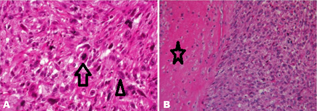
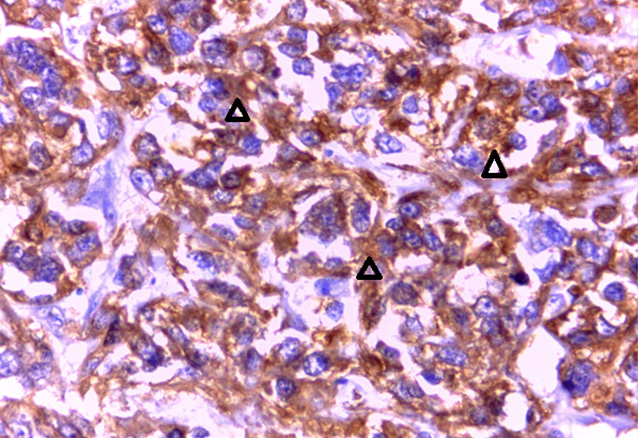
It represented the most frequent histological type with seven cases (41%) (Figure 1). The average age was 60.8 years with extremes of 47 and 80 years. Six (6) women were menopausal. The leiomyosarcoma (LMS) was graded in five patients. The tumor size was specified in four patients. It was, on average, 18.5 cm with extremes of 6 and 29 cm. In four patients, the myometrial infiltration involved 2/3 of the myometrium and the entire wall in one case. The tumors were classified as grade I for one patient, grade II for two patients, and grade III for two patients. The results of the immunohistochemical study had been presented separately for each antibody; three (three) patients received an AML immunolabeling with AML only which was positive (Figure 2); one (1) other patient was AML +; h-caldesmon +, Desmin - and myogenic -.
Endometrial stromal sarcoma
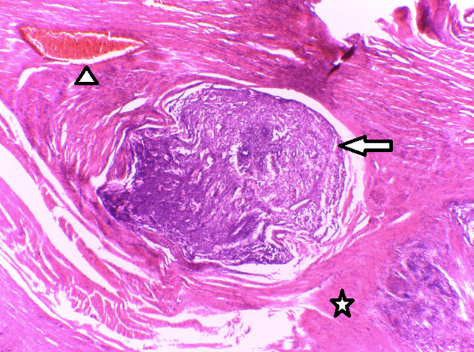
This histological type was found in five patients (30% of cases). The average age was 48.6 years with extremes of 48 and 66 years. Four (4) patients were menopausal. The tumor size was specified in three patients. It averaged 6 cm, with extremes of 3.5 and 10 cm. The invasion depth of the myometrium was mentioned in four patients (Figure 3). In three cases, the infiltration concerned the entire myometrium, and in one (1) case it concerned its internal 2/3. The histological grade showed a low grade ESS in four patients and an undifferentiated ESS in one patient. Vascular invasion was absent in three cases of low grade ESS and present in the case of the undifferentiated ESS. The immunohistochemical profile had been performed on two (2) patients, and it showed CD10+ and CD117- on a low grade ESS and CD10 +, Vimentin -, AML -, RE -, RP - on the undifferentiated ESS.
Carcinosarcoma
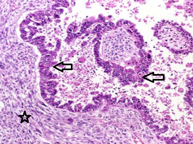
It occupied third place in uterine sarcoma in our cohort with four cases (23%) (Figure 4). The average age was 50.25 years with extremes of 14 and 77 years. Three patients were menopausal, and one (1) patient was 14 years old. The average size of the tumor was 14 cm with extremes of 8 and 12 cm. The tumor invaded the internal 2/3 of the myometrium in three patients, and the entire myometrium in one (1) patient. Carcinosarcomas were immediately labeled as highgrade tumors. The vascular invasion was positive in one case. The immunohistochemical examination had not been performed for these patients.
Adenosarcoma
It was found in one (1) 45-year-old menopausal woman. The tumor measured 5 cm on the long axis. The myometrial infiltration had not been evoked on the pathology report. The presence or absence of vascular emboli was not mentioned. A panel using CK and CD10 was positive for both markers.
DISCUSSION
Epidemiological
Considering the small size of our sampling, to numerous studies conducted in Morocco [2] and in Nigeria [3] it can be said that uterine sarcomas are relatively rare. This could be explained by the inaccessibility of archives from private laboratories in our country, hence the importance of setting a national cancer registry. In addition, a large number of samples are sent to laboratories abroad, and the results are not always available. Another reason could be related to socioeconomic difficulties in our regions. Indeed most of our patients often cannot afford the cost of treatment and pathological examination, and thus, do not have access to the care they need.
Several authors have investigated the racial distribution of sarcomas. They reported a predominance of tumors with black people which is three times higher than with white people [4]. We could not observe this difference because all our patients were black.
Histopronostic aspects
For all our patients the diagnostic of sarcoma is done to morphology. But there are several vareties of sarcoma and it is necessary to do the immunohistochemical study to give the histological type. So it is for a prognostic and epidemiological interest.
Leiomyosarcoma
The LMS is the most common histological type in our series. The average age of onset of the disease in our series is 60.8 years, well above the figures reported by most authors who found an average age around 50 [5]. The histopronostic criteria based essentially on the morphological criteria of LMSs have evolved considerably in the literature, and remain inaccurately defined. They were initially exclusively based on the mitotic activity, on the presence or absence of cytonuclearatypia and of tumor necrosis. Tumor size is currently one of the most important prognostic factors in the treatment of the LMS of the uterine. According to the WHO and according to many studies, tumors smaller than 5 cm seem to be a factor of good prognosis [6]. Other authors found a correlation between prognosis, myometrial invasion, and the presence or absence of vascular emboli. According to the latter, a lack of vascular invasion and a low myometrial invasion are factors of good prognosis [7]. On an immunohistochemical perspective, the positivity of the AML at immunohistochemistry for certain diagnosis was in agreement with other studies in which all cases were positive [8]. Immunolabeling with desmin and h-caldesmon was variable according to the samplings [9].
Endometrial stromal sarcoma
This histological type accounted for 30% of uterine sarcomas in our samplings. According to many publications, prognostic factors remain controversial. Apart from traditional prognostic factors such as the mitotic index, nuclear pleomorphism, and tumorous necrosis, other factors such as age – more than 50-yearold, black race, the tumor size, the myometrial invasion, and the negative profile of estrogen and progesterone receptors seem to be independent prognostic factors for low survival [10]. The immunohistochemistry increasingly practiced in recent years for these lesions, not only differentiates ESS from smooth muscle tumors, but also objectively and reproducibly assess the prognosis of ESSs [11]. In our sampling as in the authors’, CD10 positivity was observed in ESS cases with rates varying between 85 and 100% [11]. The negativity of hormone receptors RE and RP, as is the case in one of our patients, seems to be the result of inaccurate prognosis [11].
Carcinosarcoma
This histological type was rarely represented. It was estimated at 23% of uterine sarcomas in our cohort. Higher frequencies were also noted in South Africa [12] and France [13]. Uterine CSs are tumors diagnosed most often after 60 years of age. However, there are rare cases of uterine CS in young women [14] as is the case in our sampling in which the youngest patient was 14 years old. The histological diagnosis is easy in classical forms showing a malignant glandular epithelial contingent with or without endometrioid differentiation, and a homologous or heterologous malignant mesenchymal contingent. The homologous type allows for a better prognosis for the CS [15]. Immunohistochemistry is not essential for diagnosis; it however shows an epithelial contingent that is generally positive for anti-cytokeratin antibodies, and a positive mesenchymal contingent for vimentin.
Adenosarcoma
The frequency of adenosarcomas in our sampling was 6% of all uterine sarcomas. The ASs occur between the ages of 15 and 90 [16], as is the case in our sampling where the patient was 45-year-old. Adenosacromas are generally considered as low-grade malignancy tumors with good prognosis [16]. The immunohistochemistry has been of great help in the differential diagnosis in the absence of dense cellularity of stroma and pronounced cytonuclearatypia [16].
CONCLUSION
Uterine sarcomas are characterized by great anatomopathological heterogeneity. Immunohistochemistry is essential for positive diagnosis, for differential diagnosis and, in some cases, it allows to assess the prognosis. It is of paramount importance to reinforce the technical platform of the various pathology anatomy and cytology laboratories by implementing immunohistochemistry for a better characterization of these tumors.
REFERENCES
1.
Acharya S, Hensley ML, Montag AC, Fleming GF. Rare uterine cancers. Lancet Oncol 2005;6(12):961–71. [CrossRef]
[Pubmed]

2.
Bennani O, Himmi A, Laghzaoui M, Aderdour M. Uterine sarcoma. Apropos of 25 cases. [Article in French]. Rev Fr Gynecol Obstet 1995;90(1):12–6.
[Pubmed]
3.
Akhiwu W, Ochuba C, Okonkwo CA. Clinico-pathological features of uterine sarcomas at the university of Benin Teaching Hospital, Benin-city, Nigeria. Mary Slessor Journal of Medecine 2010;10(2).
4.
Sherman ME, Devesa SS. Analysis of racial differences in incidence, survival, and mortality for malignant tumors of the uterine corpus. Cancer 2003;98(1):176-86. [CrossRef]
[Pubmed]
5.
Horo AG, Aka KE, Fomba M, et al. Uterine inversion: An uncommon progressive forme of leiomyosarcoma uterine: Case report and review of literature. Open Journal of Obstetrics and Gynecology 2016;6(2):144–8. [CrossRef]
6.
Tavassoli FA, Devilee P. World Health Organization Classification of Tumours. Pathology and Genetics Tumours of the Breast and Female Genital Organs. Lyon: IARC Press; 2003. p. 233–44.
7.
Razafintsalama T, Leveque J, Le Gall F, et al. Uterine leiomyosarcoma. Nine case reports, review of the literature. [Article in French]. J Gynecol Obstet Biol Reprod (Paris) 1997;26(3):256–62.
[Pubmed]
8.
Farah-Klibi F, Ben Hamouda S, Ben Ramdhane S, et al. Immunohistochemical study of endometrial stromal sarcoma and smooth-muscle tumors of the uterus. [Article in French]. J Gynecol Obstet Biol Reprod (Paris) 2008;37(5):457–62. [CrossRef]
[Pubmed]
9.
Rush DS, Tan JY, Baergen RN, Soslow RA. h-Caldesmon, a novel smooth muscle-specific antibody, distinguishes between cellular leiomyoma and endometrial stromal sarcoma. Am J Surg Pathol 2001;25(2):253–8.
[Pubmed]
10.
Yoon A, Park JY, Park JY, et al. Prognostic factors and outcomes in endometrial stromal sarcoma with the 2009 FIGO staging system: A multicenter review of 114 cases. Gynecol Oncol 2014;132(1):70–5. [CrossRef]
[Pubmed]
11.
Zhu XQ, Shi YF, Cheng XD, Zhao CL, Wu YZ. Immunohistochemical markers in differential diagnosis of endometrial stromal sarcoma and cellular leiomyoma. Gynecol Oncol 2004;92(1):71–9. [CrossRef]
[Pubmed]
12.
Amant F, Dreyer L, Makin J, Vergote I, Lindeque BG. Uterine sarcomas in South African black women: A clinicopathologic study with ethnic considerations. Eur J Gynaecol Oncol 2001;22(3):194–200.
[Pubmed]
13.
Hassini A, Khemiri B, Sfar E, Chelly D, Chennoufi MB, Chelly H. Uterine sarcomas: Clinical and therapeutic aspects (10 cases). [Article in French]. J Gynecol Obstet Biol Reprod (Paris) 2006;35(4):348–55.
[Pubmed]
14.
Park JY, Kim DY, Suh DS, et al. Prognostic factors and treatment outcomes of patients with uterine sarcoma: Analysis of 127 patients at a single institution, 1989–2007. J Cancer Res Clin Oncol 2008;134(12):1277–87. [CrossRef]
[Pubmed]
15.
Inthasorn P, Carter J, Valmadre J, Beale P, Russell P, Dalrymple C. Analysis of clinicopathologic factors in malignant mixed Müllerian tumors of the uterine corpus. Int J Gynecol Cancer 2002;12(4):348–53.
[Pubmed]
16.
Sassi S, Hamdane MM, Sellami Dhouib R, et al. A rare uterine tumor in a 17 year-old girl. [Article in French]. Tunis Med 2012;90(1):881–90.
[Pubmed]
SUPPORTING INFORMATION
Author Contributions
Tonleu Linda Bentefouet - Acquisition of data, Analysis of data, Drafting the work, Revising the work critically for important intellectual content, Final approval of the version to be published, Agree to be accountable for all aspects of the work in ensuring that questions related to the accuracy or integrity of any part of the work are appropriately investigated and resolved.
Abdoul Magib Gaye - Conception of the work, Design of the work, Acquisition of data, Drafting the work, Revising the work critically for important intellectual content, Final approval of the version to be published, Agree to be accountable for all aspects of the work in ensuring that questions related to the accuracy or integrity of any part of the work are appropriately investigated and resolved.
Ibou Thiam - Conception of the work, Design of the work, Drafting the work, Revising the work critically for important intellectual content, Final approval of the version to be published, Agree to be accountable for all aspects of the work in ensuring that questions related to the accuracy or integrity of any part of the work are appropriately investigated and resolved.
Hatim J - Acquisition of data, Analysis of data, Drafting the work, Final approval of the version to be published, Agree to be accountable for all aspects of the work in ensuring that questions related to the accuracy or integrity of any part of the work are appropriately investigated and resolved.
Source of SupportNone
Data AvailabilityAll relevant data are within the paper and its Supporting Information files.
Conflict of InterestAuthors declare no conflict of interest.
Copyright© 2019 Tonleu Linda Bentefouet et al. This article is distributed under the terms of Creative Commons Attribution License which permits unrestricted use, distribution and reproduction in any medium provided the original author(s) and original publisher are properly credited. Please see the copyright policy on the journal website for more information.


