| Table of Contents | |
|
Clinical Image
| ||||||
| Herlyn-Werner-Wunderlich syndrome (OHVIRA) with recurrent haematocolpos after aspiration drainage of the obstructed hemivagina | ||||||
| Ahmad Shuib Yahaya1, Anizah Aishah Rosli1, Nik Ahmad Zuky Nik Lah2 | ||||||
|
1Specialist Trainee, Obstetric & Gynaecology Department, School of Medical Sciences, Universiti Sains Malaysia, Malaysia.
2Senior Lecturer, Obstetric & Gynaecology Department, School of Medical Sciences, Universiti Sains Malaysia, Malaysia. | ||||||
| ||||||
|
[HTML Abstract]
[PDF Full Text]
[Print This Article]
[Similar article in Pumed] [Similar article in Google Scholar] |
| How to cite this article |
| Yahaya AS, Rosli AA, Lah NAZN. Herlyn-Werner- Wunderlich syndrome (OHVIRA) with recurrent haematocolpos after aspiration drainage of the obstructed hemivagina. Edorium J Gynecol Obstet 2015;1:19–21. |
|
Case Report
| ||||||
|
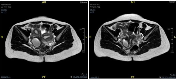
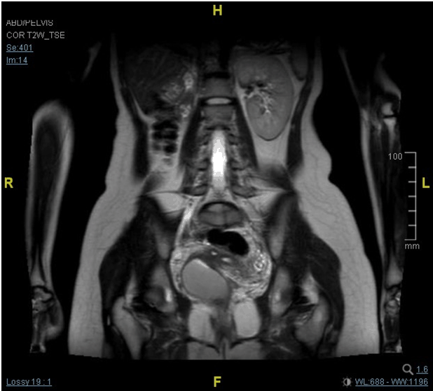
A 20-year-old single lady with abnormal menses and severe dysmenorrhea and initial assessment revealed uterine didelphys with pelvic collection suspicious of hematocolpos. There was no specific finding in the physical examination. The per rectal examination revealed extra luminal mass from the right vaginal wall with severe tenderness elicited. Abdominal ultrasonographic examination revealed the presence of two uteri with separated endometrium each. The measurement of the right uterus was 4.6x2.7 cm with endometrial thickness of 5.7 mm. There was a hypoechoic area within the vagina measuring 6.8x3.8 cm likely to be hematocolpos. The left uterus was normal, 4.4x3.8 cm, with endometrium thickness of 5 mm. Diagnostic laparoscopy and examination under anesthesia was performed and transvaginal aspiration drainage of hematocolpos was done in the same setting. 60 ml of chocolate-colored fluid was drained. The patient was symptom-free for three months and recurrence of symptomatic hematocolpos was detected during the follow-up visit. Magnetic resonance imaging (MRI) scan of pelvis was done which showed uterine didelphys with normal left uterus and enlarged right uterus with large proteinaceous fluid within the cervix with rounded pouch distally. Both of the uterine body was connected with different cervical canal but shared the same vaginal canal (Figure 1). There was an absent of the right kidney (Figure 2). Features of the MRI scan are in keeping of uterine didelphys with obstructed right hemivagina and right renal agenesis; suggestive of Herlyn-Werner-Wunderlich syndrome. Definitive treatment was planned and resection of the vaginal septum was performed. The septum was resected and the edges of the vaginal epithelium were sutured to prevent development of obstructing tissues during wound healing. Upon the follow-up visits at the clinic, the patient was asymptomatic and the examination revealed healed, unobstructed vaginal canal with no recurrence of hematocolpos. | ||||||
|
| ||||||
|
| ||||||
| ||||||
|
Discussion
| ||||||
|
Herlyn- Werner-Wunderlich syndrome or OHVIRA; (Obstructed Hemi Vagina with Ipsilateral Renal Agenesis) is a relatively rare type of Mullerian anomalies that was first described in 1922. This anomaly is generally observed in post-menarche adolescents and young women with irregular menses, dysmenorrhea, abdominal pain, and pelvic mass [1] [2]. The syndrome may be difficult to be recognized due to the infrequency of this syndrome, therefore a high index of suspicion is required. The diagnosis is often delayed as initially regular menstrual flow would be observed from unobstructed vaginal canal. Early detection is important as immediate surgical resection of the obstructed hemivagina, as to prevent complications and preserve future fertility. Among the recognized complications related to chronic cryptomenorrhea include endometriosis (delayed cases as consequence of retrograde menstruation) or pelvic adhesions and infectious collections (hematometra or pyometra and hematosalpinx or pyosalpinx). Ultrasound and MRI are widely and effectively used in the diagnosis of genitourinary anomalies. Accuracy reported from MRI scan being 100% due to its ability to elaborate uterovaginal anatomy in detail [3]. Total resection of the septum (septectomy) and marsupialization are the optimal management options. The septum should be excised as much as possible, followed by suturing the edges or marsupialization to allow drainage of the menstrual fluid and prevent recurrence of obstruction. Pregnancies had been reported before in patients with this syndrome, including from uterus ipsilateral to the obstructed vagina after resection of the septum performed [4]. | ||||||
|
Conclusion
| ||||||
|
Imaging technique is important in diagnosis of Mullerian anomalies. In case of Herlyn-Werner-Wunderlich syndrome (OHVIRA), resection of the obstructing septum is the optimal management, as compared to drainage of the haematocolpos. Keywords: Haematocolpos, Herlyn-Werner-Wunderlich, Mullerian duct anomaly, Renal agenesis | ||||||
|
Acknowledgements
| ||||||
|
Assistant Prof. Dr. Shah Reza Johan Nor | ||||||
|
References
| ||||||
| ||||||
|
SUGGESTED READING
| ||||||
| ||||||
|
[HTML Abstract]
[PDF Full Text]
|
|
Author Contributions
Ahmad Shuib Yahaya – Substantial contributions to conception and design, Drafting the article, Revising it critically for important intellectual content, Final approval of the version to be published Anizah Aishah Rosli – Substantial contributions to conception and design, Drafting the article, Revising it critically for important intellectual content, Final approval of the version to be published Nik Ahmad Zuky Nik Lah – Substantial contributions to conception and design, Drafting the article, Revising it critically for important intellectual content, Final approval of the version to be published |
|
Guarantor of submission
The corresponding author is the guarantor of submission. |
|
Source of support
None |
|
Conflict of interest
Authors declare no conflict of interest. |
|
Copyright
© 2015 Ahmad Shuib Yahaya et al. This article is distributed under the terms of Creative Commons Attribution License which permits unrestricted use, distribution and reproduction in any medium provided the original author(s) and original publisher are properly credited. Please see the copyright policy on the journal website for more information. |
|
|